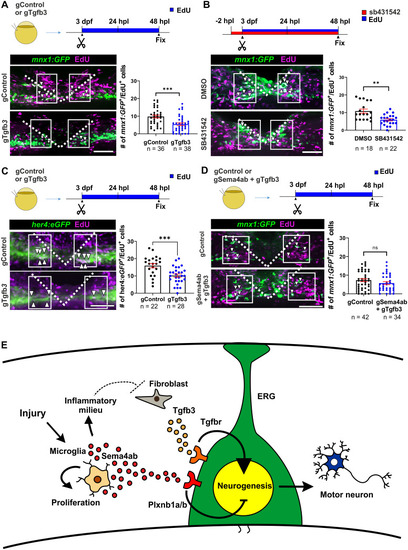
Fibroblasts promote regenerative neurogenesis via tgfb3.
(A) Somatic mutants for tgfb3 show a reduction in the number of newly generated motor neurons (gControl: 9.8 cells per larva ± 0.8; gTgfb3: 5.5 cells per larva ± 0.7; Mann–Whitney U test: p = 0.0001). (B) SB431542 treatment reduces regenerative neurogenesis (DMSO: 10.8 cells per larva ± 1.15; SB431542: 5.9 cells per larva ± 0.63; Mann–Whitney U test: p = 0.0011). (C) Lesion-induced proliferation of ERGs (her4.1:EGFP+/EdU+) is decreased in somatic mutants for tgfb3 (gControl: 15.91 cells per larva ± 1.2; gTgfb3: 10.18 cells per larva ± 0.9; Unpaired t test: p = 0.0004). (D) Double haCR injection against sema4ab and tgfb3 restores control levels of regenerative neurogenesis (gControl: 8.1 cells per larva ± 0.7; gSema4ab+gTgfb3: 6.6 cells per larva ± 0.8; Mann–Whitney U test: p = 0.1523). (E) A graphical summary of proposed interactions of sema4ab during spinal cord regeneration is shown. After spinal injury, microglia likely control neurogenesis directly by signaling through Sema4ab and Plxnb1a/b on ERGs, and indirectly by changing the injury site environment, which leads to lower expression of tgfb3, a positive regulator of regenerative neurogenesis, in fibroblasts. Additionally, Sema4ab controls microglia number by promoting cell proliferation. Error bars show SEM. Dotted lines show the injury site. Scale bars: 50 µm. Data files for graphs available in S2 Data.
|
