FIGURE

Fig 1

ID
ZDB-FIG-260629-128
Publication
Docampo-Seara et al., 2026 - The microglia-derived protein sema4ab attenuates regenerative neurogenesis after spinal cord injury in zebrafish
Other Figures
All Figure Page
Back to All Figure Page
Fig 1

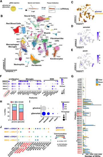
scRNA-seq indicates changes gene expression after sema4ab ablation and predicts changes in regenerative neurogenesis.
(A) A schematic of the experimental design for scRNA-seq in somatic mutants is shown. (B) Cluster analysis identifies 44 cell clusters that represent the main expected cell types in the injury site at 24 hpl. (C) A UMAP shows no major changes in cell type representation resulting from sema4ab disruption (gControl in blue, gSema4ab in yellow). (D,E) Feature plots for sema4ab (D) and apoeb (microglia; E) show overlap (black circles). (F) A dot plot of the four BDMs/microglia clusters showing that sema4ab-enriched clusters align with the expression of microglia marker genes. (G) A graph indicating differential expression of genes (DEG) between samples is shown. Cell clusters with major changes (>300 DEGs) are indicated in red. Microglia clusters MM#1 and MM#2 are boxed. (H) Graph shows the proportions of ERGs in each cell cycle phase in control gRNA-injected larvae and sema4ab gRNA-injected larvae. Note that the proportion of cells in M/G2 phase is increased in sema4ab somatic mutants (gControl: 16% vs gSema4ab: 28%). (I) Dot plot showing an increase in differentiation marker gene expression in ERG progenitors when sema4ab is disrupted. (J) Dot plot illustrating the communication probabilities of all ligand-receptor interactions identified between MM#1 and ERGs, as well as between MM#2 and ERGs. The absence of dots indicates that no interaction was detected. For a key to cluster abbreviations in B and G, see S5 Table.

Expression Data

Expression Detail
Antibody Labeling
Phenotype Data

Phenotype Detail
Acknowledgments
This image is the copyrighted work of the attributed author or publisher, and ZFIN has permission only to display this image to its users. Additional permissions should be obtained from the applicable author or publisher of the image. Full text @ PLoS Biol.