|
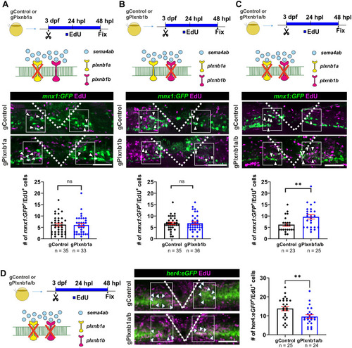
Combined plxnb1a and plxnb1b loss of function increases regenerative neurogenesis.
Photomicrographs are oriented as in Fig 2. (A,B) Regenerative neurogenesis is not changed in single somatic mutants for plxnb1a (A, gControl: 6.2 cells per larva ± 0.66; gPlxnb1a: 6.1 cells per larva ± 0.71; Mann–Whitney U test: p = 0.8371) and plxnb1b (B, gControl: 6.8 cells per larva ± 0.52; gPlxnb1b: 6.9 cells per larva ± 0.60; Mann–Whitney U test: p = 0.6511). (C) Regenerative neurogenesis is increased in double somatic mutants of plxnb1a and plxnb1b (gControl: 6.2 cells per larva ± 0.71 cells per larva; gPlxnb1a/b: 9.6 cells per larva ± 0.96; Mann–Whitney U test: p = 0.0057). (D) Lesion-induced proliferation of ERGs (her4.1:EGFP+/EdU+) is decreased in double somatic mutants for plxnb1a/b (gControl: 5.4 cells per larva ± 0.9; gSema4ab: 10.77 cells per larva ± 1.8; Mann–Whitney U test: p = 0.0219). Error bars show SEM. Scale bars: 50 µm. Data files for graphs available in S2 Data.
|