FIGURE

Fig S8

ID
ZDB-FIG-260821-31
Publication
Docampo-Seara et al., 2026 - The microglia-derived protein sema4ab attenuates regenerative neurogenesis after spinal cord injury in zebrafish
Other Figures
All Figure Page
Back to All Figure Page
Fig S8

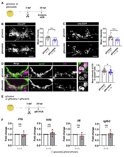
Loss of sema4ab reduces il1b and tnfa expression in vivo, but plxnb1a/plxnb1b disruption does not alter the cytokine profile of the lesion site.
(A) A schematic indicating the experimental design for cytokine quantification in situ is shown. (B) il1b:GFP fluorescence intensity reported in transgenic fish is decreased in lesioned sema4ab somatic mutants compared to control gRNA-injected fish at 24 hpl (−1.45 fold-change; Unpaired t test: p = 0.0004). (C) tnfa:EGFP fluorescence intensity reported in transgenic animals is decreased in lesioned sema4ab somatic mutants compared to control injected fish at 24 hpl (−1.49 fold-change; Unpaired t test: p < 0.0001). (D) il1b expression in microglia (apoeb+ cells) detected by HCR shows a reduction of 11% in mean intensity (Unpaired t test: p = 0.0380). (E) A schematic indicating the experimental design for qRT-PCR. (F) qRT-PCR analyses of major pro-inflammatory cytokines (il1b, tnfa, il6) and tgfb3 in plxnb1a/plxnb1b double somatic mutants show no differences to injured control gRNA-injected larvae at 24 hpl (One-sample t test; il1b: p = 0.537; tnfa: p = 0.4840; il6: p = 0.5464; tgfb3: p = 0.687). Each dot for qRT-PCR represents a pool of 50 larvae. β-actin was used as housekeeping gene. Raw data for qRT-PCR can be found in S5 and S6 Data. Error bars show SEM. Scale bars: 50 µm. Data files for graphs available in S3 Data.

Expression Data

Expression Detail
Antibody Labeling
Phenotype Data

Phenotype Detail
Acknowledgments
This image is the copyrighted work of the attributed author or publisher, and ZFIN has permission only to display this image to its users. Additional permissions should be obtained from the applicable author or publisher of the image. Full text @ PLoS Biol.