
Fig 2
- ID
- ZDB-IMAGE-260629-134
- Publication
- Docampo-Seara et al., 2026 - The microglia-derived protein sema4ab attenuates regenerative neurogenesis after spinal cord injury in zebrafish
- All Figures
- Figures for Docampo-Seara et al., 2026
|
Fig 2
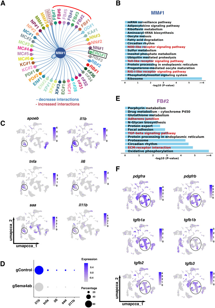
scRNA-seq indicates changes in cell-cell interactions between microglia and other cell types after sema4ab ablation.
(A) CellChat analysis for MM#1 shows interactions with all cell types and a general decrease in these interactions (blue lines) when sema4ab is disrupted. Interactions of MM#1 with ERGs (black box), fibroblasts (red curved line) and MM#2 (pink box) are highlighted. (B) KEGG analysis of the main cluster that expresses sema4ab (MM#1) shows changes in immune activation pathways (highlighted in red). (C) Feature plots show enrichment of pro-inflammatory cytokines and inflammatory markers (il1b, tnfa, il6, saa, il11b) in microglia (apoeb, circle). (D) Dot plot showing reduction of expression of pro-inflammatory cytokines and inflammatory markers after sema4ab ablation. (E) KEGG analysis of the signaling pathways most affected by sema4ab disruption in fibroblast-like cell cluster FB#2 is shown. tgfb signaling and ECM interaction-related terms are highlighted in red. (F) Feature plots showing enrichment of anti-inflammatory cytokines (tgfb1a, tgfb1b, tgfb2, tgfb3) in fibroblasts (pdgfra, pdgfrb; circled).