|
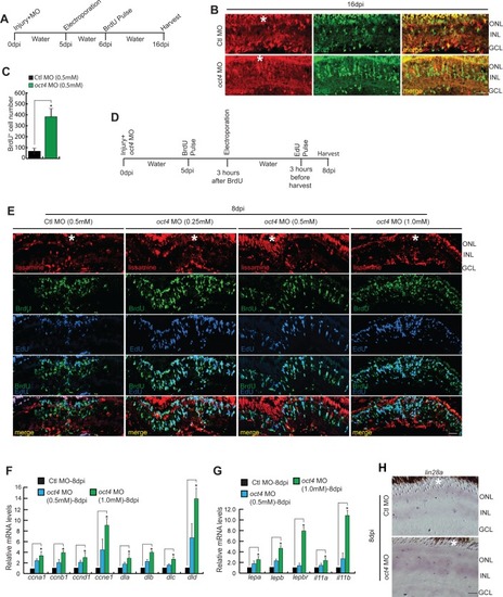
Effects of <italic>oct4</italic> late knockdown on cell proliferation and gene expression during retina regeneration.(A) An experimental timeline that describes the injury, MO delivery, electroporation, BrdU pulse, and retina harvest at 16 dpi. (B, C) IF confocal microscopy images of retinal cross sections show increased BrdU+ MGPCs at 16 dpi in oct4 knockdown retina from fifth day onwards (B), which is quantified (C); *P < 0.007 (t test), N = 4. (D) An experimental timeline that describes the injury, MO delivery, BrdU pulse, electroporation, and retina harvest after EdU pulse at 8 dpi. (E) IF confocal microscopy images of retinal cross sections show increased BrdU+ MGPCs at 8 dpi in oct4 knockdown retina from fifth day onwards. (F, G) qPCR analysis reveals the increase in mRNA levels of cyclins and delta genes (F), *P < 0.03 (t test), N = 4, along with cytokines (G), *P < 0.04 (t test), N = 4, in late oct4 knockdown retina at 8 dpi. (H) BF microscopy images of retinal cross sections show increased lin28a levels with late oct4 knockdown at 8 dpi. Ctl MO is control MO. Error bars are SD. (B, E, H) Scale bars, 10 μm (B, E, H); the asterisk marks the injury site in (B, E, H). GCL, ganglion cell layer; INL, inner nuclear layer; ONL, outer nuclear layer (B, E, H).
|