|
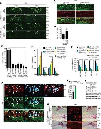
Oct4-influenced gene expression and cell proliferation during retinal regeneration.(A, B) IF confocal microscopy images of 4 dpi retinal cross sections show BrdU+ MGPCs in zeb2a mRNA-transfected conditions along with gfp mRNA-transfected control retina, with ctl MO and oct4 MO-electroporated conditions (A), which are quantified (B); *P < 0.001 (t test), N = 4. (C, D) IF confocal microscopy images of 2 dpi retinal cross sections show PCNA+ MGPCs in hdac1 mRNA-transfected conditions along with gfp mRNA-transfected control retina (C), which is quantified (D). (E, F) qPCR analysis reveals the variations in mRNA of zeb (E), *P < 0.04 (t test), N = 4, genes, along with miR-200a, miR-200b, miR-143, and miR-145 (F), *P < 0.005 (t test), N = 4, in oct4 mRNA-transfected retina, at 2 dpi. (G) IF confocal microscopy images of retinal cross section show double-FISH of oct4 and tgfbi mRNA in 4 dpi retina. White arrowheads mark EdU+ cells and white arrows mark oct4+/tgfbi+ cells. (H, I) BF and IF microscopy images of retinal cross sections (H) and qPCR (I) show increased ascl1a and decreased BrdU with Oct4 overexpression at 2 dpi. (J) Western blot analysis of Ascl1a, PCNA, and GFP compared with Gapd from oct4 mRNA-transfected retinal extracts at 2 dpi. Ctl MO is control MO. Error bars are SD. (A, C, G, H) Scale bars, 10 μm; the asterisk marks the injury site, GCL, ganglion cell layer; INL, inner nuclear layer; ONL, outer nuclear layer (A, C, G, H).
|