
Zebrafish Anatomical Dictionary
Structure description: sensory patches of the ear
by Tanya Whitfield
Name: sensory patches of the ear
Abbreviation: : am, pm, anterior and posterior maculae; ac, lc, pc, anterior, lateral and posterior cristae
Synonyms: anterior macula, posterior macula (also known as the posteromedial macula), cristae
Figures:
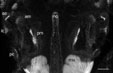
 4
day ear
4
day ear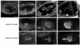 Sensory patches
 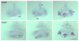 bmp4/msx expression
 |
 Dissected ears
 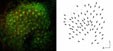 Anterior macula polarities Anterior macula polarities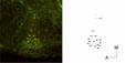 Lateral crista polarities
 |
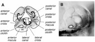
Description: Patches of thickened, pseudostratified epithelium of the inner ear, consisting of regular arrays of sensory hair cells interspersed with supporting cells. Each patch has its own charcteristic shape and polarity pattern. The anterior macula is rounded. At 5 days of development, the hair cell polarity pattern of the anterior macula resembles that of the adult utricle (Platt, 1993), with most hair cells radiating out laterally from a medial point, and a rim of hair cells around the lateral edge of the macula pointing medially. The day 5 posterior macula has a rounded posterior section and a slim anterior projection. At this stage, in the anterior projection, hair cells are arranged in an antiparallel fashion, while in the posterior region of the macula, hair cells point away from a midline separating dorsal and ventral halves. This is the "standard" four-quadrant pattern for a teleost sacculus (Popper et al., 1982), but the adult zebrafish sacculus has been described to have a "vertical" pattern, i.e. lacking the anterior region of antiparallel organisation (Platt, 1993). The anterior and posterior cristae are saddle shaped; the lateral crista, at day 4 and 5, is triangular, with a few hair cells separated from, and lying medial to, the apex of the triangle. Hair cells within a crista all point in the same direction.
Homologues:
- Human: sensory patches of the inner ear
- Mouse: sensory patches of the inner ear
- Chicken: sensory patches of the inner ear
- Frog: sensory patches of the inner ear
- Fly: none
Stages:
- First appears at: 24h (differentiated ear hair cells; the decision to become sensory epithelium probably occurs at a much earlier stage)
- Disappears (or changes name) at: Unknown
Parents (forms from): cranial placodal ectoderm (otic placode)
Children:
- Presumptive (thought to give rise to): The anterior macula of the larva corresponds to the utricular macula of the adult; the posterior macula probably forms the saccular macula. The origins of the lagena and macula neglecta of the adult are unknown.
- Anlage (known to give rise to): Unknown
Group (member of):
- Anatomical (group member): Ear
- Functional (group member): Mechanosensory systems
Markers:
- mRNA:
- Antibodies:
- Other: None
Publications:
- Primary: Haddon, C., and Lewis, J. (1996). Early ear development in the embryo of the zebrafish, Danio rerio. Journal of Comparative Neurology 365, 113-123.
- Secondary:
Popper, A. N., Platt, C., and Saidel, W. M. (1982). Acoustic functions in the fish ear. Trends in Neurosciences 5, 276-280.
Comments: None