
|
Image description by: Tanya Whitfield
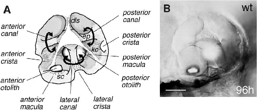
Anatomical structures shown: ear, semicircular canals, maculae and cristae, otoliths
Stage: day 4 larva
Genetic (background) strain: Tuebingen
Genotype: Tu wild-type
Animal state: live
Labeling: none; DIC optics
Description: A. Drawing of a wild-type zebrafish ear at four days of development, to give a three-dimensional impression of the structures within the vesicle. B. DIC image of an ear at the same stage, focussed on the anterior otolith. Epithelial projections (ep) within the ear form hubs of the developing semicircular canals (curved arrows). Each canal (anterior, lateral and posterior) is associated with a small sensory patch or crista, while each otolith overlies a larger sensory patch or macula. Kinocilia of crista hair cells (kc) are long, and project into the canal lumens. Kinocilia of the maculae are shorter, and the otoliths appear to sit directly on the stereociliary bundles (sc) of the macular hair cells. The smaller (anterior) otolith lies in a lateral position; the larger (posterior) otolith lies medially. A three-pronged structure is present on the lateral wall of the ear (unshaded in diagram; identity unknown). The dorsal prong of this appears to be continuous with the dorsolateral septum (dls). Scale bar, 50 µm.
Publication containing this image: T. Whitfield (unpublished).
| Preparation | Image Form | View | Direction |
| whole-mount | still | parasagittal | anterior to left |