
- Title
-
Smyd3 is required for the development of cardiac and skeletal muscle in zebrafish
- Authors
- Fujii, T., Tsunesumi, S., Yamaguchi, K., Watanabe, S., and Furukawa, Y.
- Source
- Full text @ PLoS One
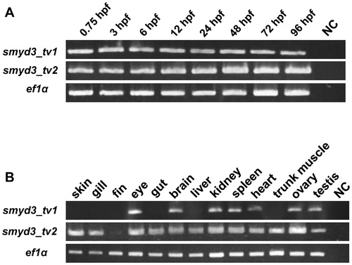
Expression of smyd3_tv1 and smyd3_tv2 in the developing zebrafish embryos and adult tissues (A) RT-PCR analysis was performed using smyd3_tv1 and smyd3_tv2-specific primer sets, with RNA extracted from zebrafish embryos at 0.75, 3, 6, 12, 24, 48, 72, and 96 hpf. NC: negative control (RNase free water). Expression of ef1α served as an internal control. (B) Expression of smyd3_tv1 and smyd3_tv2 in various adult tissues. |

Effect of Smyd3 knockdown in zebrafish embryos by Smyd3-MO or Smyd3-SB-MO. (A, B, C, and D) Suppression of smyd3 was examined at 10 hpf in embryos injected with smyd3-EGFP mRNA alone (A), smyd3-EGFP mRNA and Smyd3-mis-MO (B), and smyd3-EGFP mRNA and Smyd3-MO (C). Signals of EGFP were examined in the fluorescent macroscope (lower panel). The frequencies of EGFP-positive embryos were (A) 78.7%±2.3, (B) 83.7%±1.5, and (C) 17.7%±3.4. Data are shown as means±SEM. (D) Effect of Smyd3-SB-MO on smyd3 transcripts. The wild-type transcripts were detected as a band at 465 bp and the aberrant transcripts at 401 bp. The lower panel shows the expression of ef1α as a control. (E and F) Phenotype of embryos injected with Smyd3-MO (E) or Smyd3-mis-MO (F) at 72 hpf. The pericardial edema (arrow head) and curved trunk (arrow) were observed in Smyd3 morphants (E). (G, H, I, and J) Morphological classification of heart defect. The degree of cardiac defect in the morphants was classified into three grades at 48 hpf. Grade1: Heart shows abnormality with mild looping defect and pericardial edema (H); Grade2: Heart shows abnormality with moderate looping and defect and pericardial edema (I); Grade3: Heart shows abnormality with string-like heart with severe pericardial edema (J). Normal: Normal heart (G). Embryos are shown in lateral view. PHENOTYPE:
|
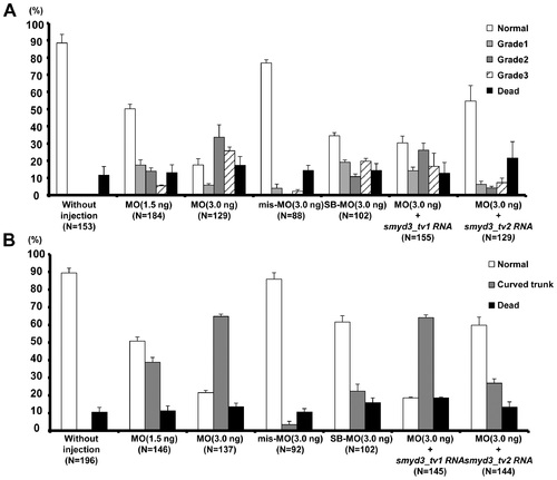
Frequencies of cardiac and trunk defects in Smyd3 morphants treated with different concentration of morpholinos, and with or without pre-injection with smyd3_tv1 or smyd3_tv2 mRNA. (A) Frequencies of heart defects in embryos injected with 1.5 or 3.0 ng of Smyd3-MO, 3.0 ng of Smyd3-mis-MO, 3.0 ng of Smyd3-SB-MO, 3.0 ng of Smyd3-MO in combination with smyd3_tv1 or smyd3_tv2 mRNA at 48 hpf. The histogram shows percentages of embryos with Normal (open box), Grade1 (light gray box), Grade2 (dark gray box), Grade3 (hatched box) or dead embryos (closed box). (B) Frequencies of curved trunk in embryos injected with 1.5 or 3.0 ng of Smyd3-MO, 3.0 ng of Smyd3-mis-MO, 3.0 ng of Smyd3-SB-MO, 3.0 ng of Smyd3-MO in combination with smyd3_tv1 or smyd3_tv2 mRNA at 48 hpf. The histogram shows percentages of embryos with Normal (open box), curved trunk (gray box), or dead embryos (closed box). Error bars represent the SEM. N is the total number of injected animals. PHENOTYPE:
|

In situ hybridization analysis of ALPM and cardiac chamber markers. (A, B, C, and D) Expression gata5, scl, nkx2.5 and hand2 in Smyd3 morphants, control embryos injected with Smyd3-mis-MO and without injection at 12 hpf. (E, F, G, H, I, and J) Expression of vmhc, amhc, and cmlc2 in the morphants, control embryos and without injection embryos at 24 hpf (E, F, and G) and 48 hpf (H, I, and J). Arrowhead indicates abnormal vmhc expression in the atrium (H). Embryos are shown in dorsal view, anterior toward the left (A, B, C, D, E, F, and G). Embryos are shown in frontal view, dorsal toward the left (H, I, and J). EXPRESSION / LABELING:
|
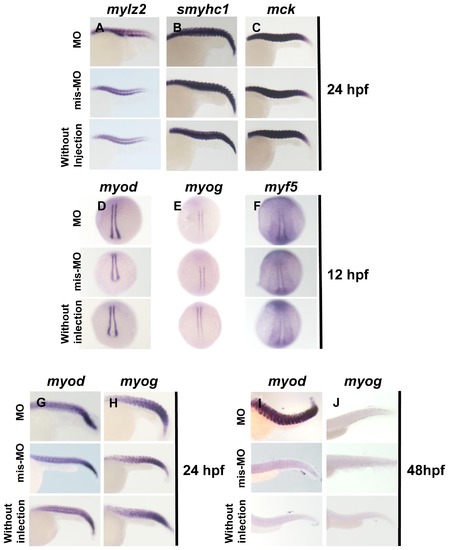
In situ hybridization analysis of terminal differentiation markers of skeletal muscle and myogenic regulatory factors. (A, B, and C) Expression of mylz2, smyhc1 and mck in Smyd3 morphants, control embryos injected with Smyd3-mis-MO and without injection at 24 hpf. (D, E, F, G, H, I, and J) Expression of myod, myog, and myf5 in Smyd3 morphants, control embryos injected with Smyd3-mis-MO and without injection at 12 hpf (D, E, and F), 24 hpf (G and H) and 48 hpf (I and J). Embryos are shown in lateral view, anterior toward the left (A, B, C, G, H, I, and J). Embryos are shown in dorsal view, anterior toward the top (D, E, and F). EXPRESSION / LABELING:
|