
- Title
-
casanova plays an early and essential role in endoderm formation in zebrafish
- Authors
- Alexander, J., Rothenberg, M., Henry, G.L., and Stainier, D.Y.R.
- Source
- Full text @ Dev. Biol.
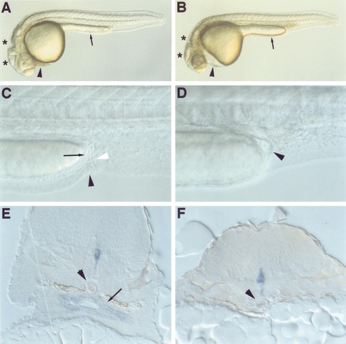
cas mutants lack a gut tube. Nomarski optical images (A–D) and histological sections (E, F) of embryos at 24 (A, B), 36 (C, D), and 48 hpf (E, F). Compared to a wild-type sibling (A), the cas mutant (B) shows pericardial edema (arrowhead), collapsed brain ventricles (asterisks), and a thicker yolk extension (arrow). In wild-type embryos (C) the still-forming intestine (arrow) and anal opening (black arrowhead) are visible, immediately anterior to the pronephric ducts (white arrowhead). Neither the intestine nor the anal opening is evident in cas mutants (D); arrowhead indicates the cyst present in cas mutants. (E) The shh-expressing gut tube (arrow) sits between the notochord (arrowhead) and the yolk in wild-type embryos. In cas mutants (F) no gut tube or shh-expressing endodermal cells are evident and the notochord (arrowhead) rests nearly upon the yolk. Expression of shh in the floor plate is evident in both wild-type and cas mutant embryos. (A–D) Lateral views, anterior to the left and dorsal to the top; (E, F) transverse sections through the upper trunk and yolk ball. |
cas mutants do not express molecular markers of endoderm differentiation. Wild-type (A, C, E) and cas mutant (B, D, F) embryos were examined at the 25-somite stage (21.5 hpf) for expression of axial (A, B), fkd7 (C, D), and gata4 (E, F). (A) axial is expressed in the ventral neuroectoderm and anterior endoderm (arrowhead); endodermal axial expression is absent from cas mutants (B). Similarly, fkd7 and gata4 are expressed in the endoderm of wild-type (arrowheads in C, E) but not cas mutant (D, F) embryos. fkd7 is also expressed in the floor plate and hypochord (C), which is posteriorly shortened in cas mutants (arrow in D). Endodermal gata4 expression defines a region of the gut tube that will form the liver. Myocardial gata4 expression (arrow in E) illustrates the cardia bifida present in cas mutants (arrows in F). (A–D) Lateral views, anterior to the left, dorsal to the top; (E, F) dorsal views, anterior to the left. EXPRESSION / LABELING:
PHENOTYPE:
|
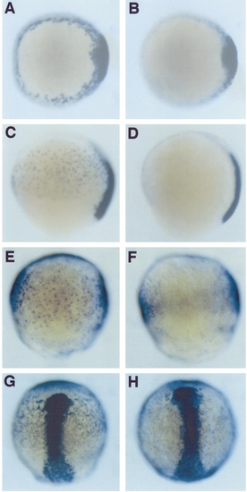
The endoderm is defective in cas mutants from the onset of gastrulation. axial (A–D), gata5 (E, F), and fkd2 (G, H) expression in wild-type (A, C, E, G) and cas (B, D, F, H) mutant embryos at shield (A, B), 80% epiboly (C–F), and 90% epiboly (G, H) stages. Soon after the onset of gastrulation (A) axial is expressed in the embryonic shield and endodermal precursors located throughout the hypoblast. Endodermal axial expression is absent in cas mutants, while expression in the embryonic shield is normal (B); the few axial-expressing cells just outside the shield in the cas mutant are likely notochord precursors that have not yet completed dorsal convergence. At midgastrulation wild-type embryos express axial in the endodermal precursors and the prechordal plate and notochord (C); no endodermal expression of axial is seen in cas mutants (D). gata5 expression also identifies endodermal precursors within the hypoblast of wild-type (E) but not cas mutant (F) embryos. gata5 expression in the anterior lateral mesoderm precursors and YSL, which is out of focus (E), appears normal in cas mutants (F). fkd2 is expressed in endodermal precursors in wild-type embryos, as well as in the YSL and axial mesoderm (G). cas mutants specifically lack endodermal fkd2 expression (H). (A, B) Animal pole views; (C, D) left lateral views, anterior to the top; (E–H) dorsal views, anterior to the top. EXPRESSION / LABELING:
PHENOTYPE:
|
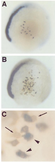
cas acts cell autonomously in the endodermal progenitors. A cas mutant host at 80% epiboly into which wild-type cells were transplanted contains several axial-expressing endodermal precursors in the lateral hypoblast (A). These cells all derive from the wild-type donor as they also contain the biotin dextran lineage tracer (brown stain in B); no mutant host cell was ever observed to form endoderm, as judged by axial expression. Under higher magnification (C) the presence of biotin dextran in the axial-expressing endodermal precursors is clearly seen (arrowheads indicate brown cells with purple cytoplasm); several cells not expressing axial also contain biotin dextran (arrows indicate brown cells). In 53 wild-type to wild-type control transplantations, we observed four cases in which transplanted cells formed axial-expressing endoderm (data not shown). Wild-type cells transplanted into cas mutant hosts formed axial-expressing endoderm in 5 of 77 cases (P = 0.5-0.9). cas mutant cells were never observed to form axial-expressing endoderm when transplanted into wild-type hosts (33 events; P < 0.1). (A, B) Right lateral views, anterior to the top; (C) high-magnification view of B. |
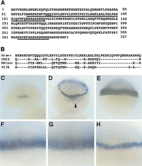
Sequence and expression of a zebrafish Mixer homologue. (A) Predicted amino acid sequence of zebrafish Mixer; the homeodomain and a C-terminal acidic domain are underlined. (B) Comparison of the homeodomains of zebrafish Mixer (Mixer), CMIX, Xenopus Mixer (XMixer), and Milk; dashes indicate conserved residues. Expression of mixer (C–F), ntl (G), and gata5 (H) in wild-type embryos at sphere (C), dome (D), and 50% epiboly (E–H) stages. mixer expression initiates in a group of cells at the dorsal margin (C), then spreads throughout the marginal zone (D) where it is maintained at the onset of gastrulation (E). mixer also appears to be expressed in the dorsal YSL at dome stage (arrowhead in D). ntl expression (G) at the onset of gastrulation encompasses essentially the same cells as does mixer expression (F), while gata5 expression (H) is limited to a subset of these cells (compare F–H). (C, D) Dorsal views; (E–H) lateral views. (F–H) High-magnification Nomarski optics images. EXPRESSION / LABELING:
|

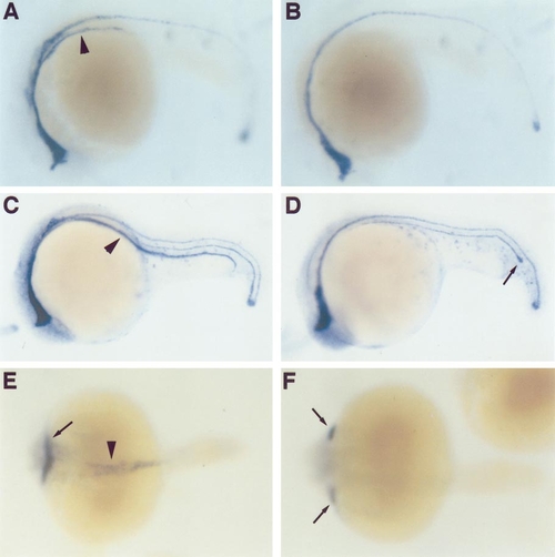
The forerunner cells do not form in cas mutants. Wildtype and cas mutant embryos were examined by light microscopy (A, B), in situ hybridization for expression of ntl (C, D), syto-11 fluorescence (E, F), and Nomarski optics (G, H). Embryos are at the following stages: (A, B) 6-somite, (C, D) 70% epiboly, (E, F) 90% epiboly, and (G, H) shield. Kupffer’s vesicle, formed by the forerunner cells (FRs), is easily seen in the tail of wild-type embryos (arrowhead in A) but not in cas mutants (B). ntl is normally expressed in the FRs (arrowhead in C) but this expression is lacking in cas mutants (D). The FR cluster (arrowhead in E) can be visualized by labeling with the fluorescent dye syto-11; cells of the enveloping layer (EVL) also take up syto-11 and thus fluoresce. cas mutants appear to lack a FR cluster (F); several syto-11-labeled EVL cells are seen. Using Nomarski optics the FRs can be directly visualized at the shield stage as a cluster of cells (arrowheads) that obscures part of the dorsal margin (arrows). No FRs are seen in a cas mutant embryo (H), allowing the margin to be easily traced (arrows). (A, B, E, F) Posterior views, dorsal to the right; (C, D, G, H) dorsal views, anterior to the top. EXPRESSION / LABELING:
PHENOTYPE:
|
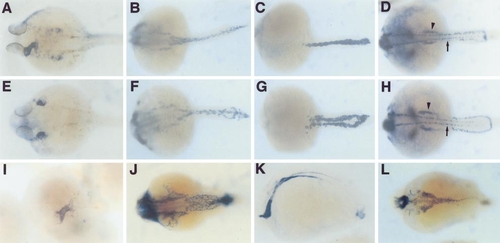
Morphogenesis of several mesodermal derivatives is defective in cas mutants. Wild-type (A, B, C, D), cas (E, F, G, H), and kny (I, J, K, L) embryos showing expression of nkx2.5 (A, E, I), tie2 (B, F, J), gata1 (C, G), pax2.1 (D, H), and axial (K, L). Embryos are at 24 (A, E, I–L) and 21.5 hpf (B–D, F–H). By 24 hpf the definitive heart tube has formed in wild-type embryos (A) while two “hearts” are evident in cas mutants (E). Pharyngeal endodermal expression of nkx2.5 is also missing in cas mutants (compare A to E). Endothelial precursors assemble smoothly in the midline of wild-type (B) but not cas mutant (F) embryos. In wild-type embryos the bilateral red blood cell precursors meet at the midline (C), but are positioned more laterally in cas mutants (G). The bilateral pronephroi (arrowheads) and pronephric ducts are also positioned more laterally in cas mutants (compare D to H); the arrows indicate the pax2.1-expressing spinal commissural interneurons. kny mutants form endoderm normally, although it is more broadly distributed (K, L); they do not exhibit cardia bifida (I); and their trunk endothelium appears smoothly although again more broadly arranged in the midline (J). All are dorsal views, anterior to the left, except (K), which is a lateral view, anterior to the left. EXPRESSION / LABELING:
PHENOTYPE:
|

Unillustrated author statements EXPRESSION / LABELING:
|
Reprinted from Developmental Biology, 215(2), Alexander, J., Rothenberg, M., Henry, G.L., and Stainier, D.Y.R., casanova plays an early and essential role in endoderm formation in zebrafish, 343-357, Copyright (1999) with permission from Elsevier. Full text @ Dev. Biol.