
- Title
-
Transforming growth factor-β receptor I kinase plays a crucial role in oligodendrocyte regeneration after demyelination
- Authors
- Lee, Y., Jung, I., Lee, D.W., Seo, Y., Kim, S., Park, H.C.
- Source
- Full text @ Biomed. Pharmacother.
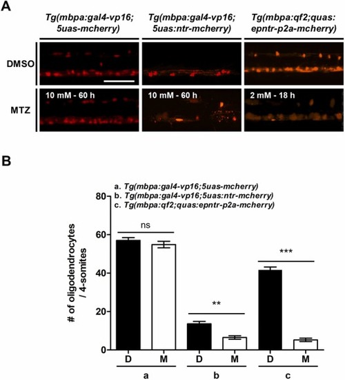
The transgenic zebrafish model in the present study has superior oligodendrocyte ablation efficiency. (A) All images are lateral views of the spinal cord of transgenic zebrafish larvae, 6 and 8 days post-fertilization (dpf), anterior to the left and dorsal to the top. (B) In the spinal cord, fluorescence indicates mbpa:mCherry+ cells (oligodendrocytes, OLs). The graph represents the number of OLs in each 4-somite area. The experiment was repeated thrice (n = 10 per group). The data are presented as the mean ± SD. P-values were calculated using an unpaired Student’s t-test. Significance is indicated by **P < 0.01 and ***P < 0.001 vs. DMSO. ns: not significant. Scale bar: 200 μm. PHENOTYPE:
|
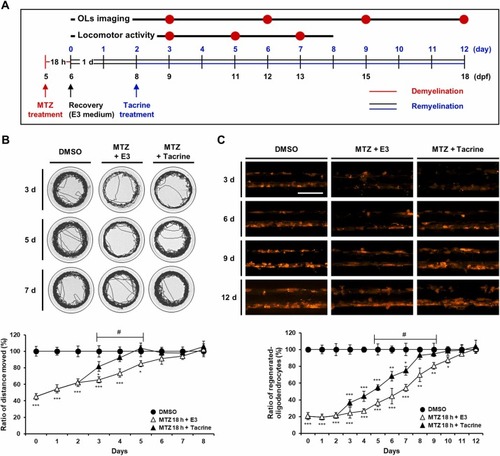
The optimal time point for screening is determined by the change in improved point achieved by tacrine treatment after demyelination. (A) Schematic diagram for demyelination and remyelination assay conditions using Tg(mbpa:qf2;quas:epntr-p2a-mcherry) zebrafish larvae. (B) Representative tracking image of a 5-min recording at 9, 11, and 13 days post-fertilization (dpf). The graph shows the ratio of the distance moved at 6–14 dpf. (C) Lateral views of the spinal cord of transgenic zebrafish larvae at 9, 12, 15, and 18 dpf, anterior to the left and dorsal to the top. In the spinal cord, fluorescence indicates mbpa+-OLs. The graph shows the ratio of the oligodendrocyte (OL) number in each 4-somite area at 6–18 dpf. The experiment was repeated thrice. The data are presented as the mean ± SD. n ≥ 10 independent experiments. P-values were calculated using one-way ANOVA followed by Tukey’s test. Significance is indicated by *P < 0.05, **P < 0.01, and ***P < 0.001 vs. DMSO; #P < 0.05 vs. MTZ 18 h + E3. Scale bar: 200 μm. PHENOTYPE:
|
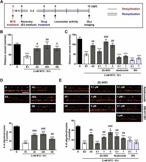
Pharmacological control of the TGF-β1 signaling pathway affects locomotor activity and oligodendrocyte regeneration. (A) Schematic representation of the treatment protocols using Tg(mbpa:qf2;quas:epntr-p2a-mcherry) zebrafish larvae. The zebrafish larvae were treated with 2 mM metronidazole (MTZ) for 18 h 5 days post-fertilization (dpf). After that, they were maintained in E3 medium and treated with AZ-12601011 (10 nM), A83–01 (10 nM), SB-505124 (50 nM), (E)-SIS3 (0.1, 1, and 5 μM), asiaticoside (0.1 and 1 μM), and SRI-011381 (0.1 and 1 μM) for 1 and 4 days. (B) Effects of TGF-βRI kinase inhibitors on distance moved 9 dpf. (C) Changes in distance moved at 9 dpf when the TGF-β signaling pathway was inhibited by pharmacological drugs. (D, E) Lateral views of the spinal cord of transgenic zebrafish larvae at 12 dpf, anterior to the left and dorsal to the top. In the spinal cord, fluorescence indicates mbpa+-OLs. The graphs show the number of oligodendrocytes (OLs) in each 4-somite area. The experiment was repeated thrice. The data are presented as the mean ± SD; n ≥ 10 independent experiments. P-values were calculated using one-way ANOVA followed by Tukey’s test. Significance is indicated by *P < 0.05, **P < 0.01, and ***P < 0.001 vs. DMSO; #P < 0.05, ##P < 0.01, and ###P < 0.001 vs. MTZ 18 h + E3. Scale bar: 200 μm. PHENOTYPE:
|
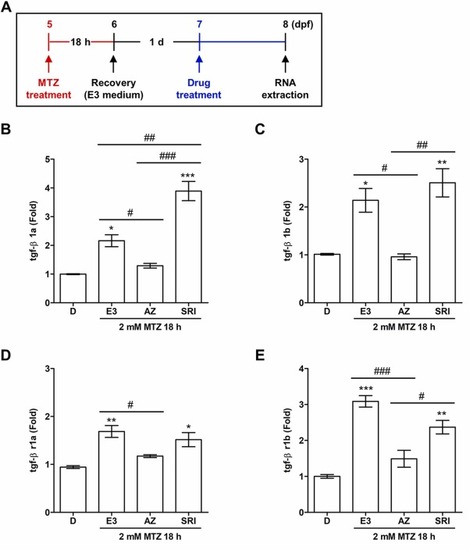
TGF-βRI kinase inhibitor downregulates TGF-β1 and TGF-βRI mRNA expression levels post-demyelination. (A) Schematic representation of the treatment protocols using Tg(mbpa:qf2;quas:epntr-p2a-mcherry) zebrafish larvae. (B-E) RT-qPCR results for tgf-β1a, tgf-β1b, tgf-βr1a, and tgf-βr1b mRNA expression. The experiment was repeated thrice. The data are presented as the mean ± SD; n ≥ 20 independent experiments. P-values were calculated using one-way ANOVA followed by Tukey’s test. Significance is indicated by *P < 0.05, **P < 0.01, and ***P < 0.001 vs. DMSO; #P < 0.05, ##P < 0.01, and ###P < 0.001 vs. MTZ 18 h + E3 or MTZ 18 h + AZ. |
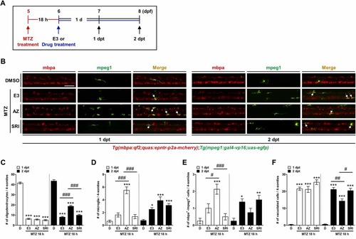
TGF-βRI kinase inhibitor increases immune responses in the early stages after oligodendrocyte ablation. (A) Schematic representation of the treatment protocols using Tg(mbpa:qf2;quas:epntr-p2a-mcherry); Tg(mpeg1:gal4-vp16;uas-egfp) zebrafish larvae. (B) Lateral views of the spinal cord of transgenic zebrafish larvae at 7 and 8 days post-fertilization (dpf), anterior to the left and dorsal to the top. In the spinal cord, fluorescence indicates mbpa+-OLs, mpeg1+-microglia, and phagocytic clearance of cellular debris by microglia (mbpa and mpeg1 double-positive cells, white allow). (C-F) Numbers of mbpa+-, mpeg1+-, mbpa+mpeg1+-, and vacuolated cells in each 4-somite area 1 and 2 days post-treatment (dpt). The experiment was repeated thrice. The data are presented as the mean ± SD; n ≥ 11 independent experiments. P-values were calculated using one-way ANOVA followed by Tukey’s test. Significance is indicated by *P < 0.05, **P < 0.01, and ***P < 0.001 vs. DMSO; #P < 0.05, ##P < 0.01, and ###P < 0.001 vs. MTZ 18 h + E3 or MTZ 18 h + AZ. Scale bar: 200 μm. PHENOTYPE:
|
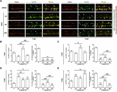
TGF-βRI kinase inhibitor increases the number of oligodendrocyte progenitor cells early after demyelination. (A) Lateral views of the spinal cord of transgenic zebrafish larvae at 1 and 4 days post-treatment (dpt), anterior to the left and dorsal to the top. In the spinal cord, fluorescence indicates sox10+-OPCs (arrowhead) and mbpa+sox10+-mature oligodendrocytes (OLs) (arrow) in each 2-somite area. (B-E) Numbers of sox10+- and mbpa+sox10+-cells in each 4-somite area 1–4 dpt. The experiment was repeated thrice. The data are presented as the mean ± SD; n ≥ 8 independent experiments. P-values were calculated using one-way ANOVA followed by Tukey’s test. Significance is indicated by *P < 0.05 and ***P < 0.001 vs. DMSO; #P < 0.05, ##P < 0.01, and ###P < 0.001 vs. MTZ 18 h + E3 or MTZ 18 h + AZ. ns: not significant. Scale bar: 200 μm. PHENOTYPE:
|
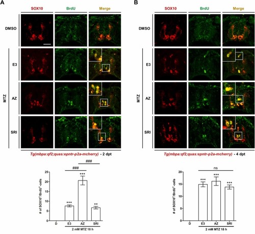
TGF-βRI kinase inhibitor promotes oligodendrocyte progenitor cell proliferation early after demyelination. Confocal images of a transverse section of the spinal cord of demyelinated zebrafish larvae at 2 and 4 days post-treatment (dpt). Sections are labeled for SOX10 and BrdU; the enlarged images show SOX10+BrdU+-cells. The graphs show the number of BrdU+-cells among the SOX10+-cells 2 and 4 dpt; the double-positive cells are counted in 10 sections per larva. The experiment was repeated thrice. The data are presented as the mean ± SD; n ≥ 8 independent experiments. P-values were calculated using one-way ANOVA followed by Tukey’s test. Significance is indicated by **P < 0.01 and ***P < 0.001 vs. DMSO; ###P < 0.001 vs. MTZ 18 h + E3 or MTZ 18 h + AZ. ns: not significant. Scale bar: 20 μm. |