
|
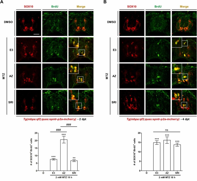
Fig. 7 TGF-βRI kinase inhibitor promotes oligodendrocyte progenitor cell proliferation early after demyelination. Confocal images of a transverse section of the spinal cord of demyelinated zebrafish larvae at 2 and 4 days post-treatment (dpt). Sections are labeled for SOX10 and BrdU; the enlarged images show SOX10+BrdU+-cells. The graphs show the number of BrdU+-cells among the SOX10+-cells 2 and 4 dpt; the double-positive cells are counted in 10 sections per larva. The experiment was repeated thrice. The data are presented as the mean ± SD; n ≥ 8 independent experiments. P-values were calculated using one-way ANOVA followed by Tukey’s test. Significance is indicated by **P < 0.01 and ***P < 0.001 vs. DMSO; ###P < 0.001 vs. MTZ 18 h + E3 or MTZ 18 h + AZ. ns: not significant. Scale bar: 20 μm.