
- Title
-
Samd7 represses short-wavelength cone genes to preserve long-wavelength cone and rod photoreceptor identity
- Authors
- Volkov, L.I., Ogawa, Y., Somjee, R., Vedder, H.E., Powell, H.E., Poria, D., Meiselman, S., Kefalov, V.J., Corbo, J.C.
- Source
- Full text @ Proc. Natl. Acad. Sci. USA

Samd7 is expressed in immature photoreceptors, red cones, green cones, and rods in the larval zebrafish retina. (A) UMAP (Uniform Manifold Approximation and Projection) of scRNA-seq data from 4-dpf crx:GFP+ photoreceptors. (B) scRNA-seq results show that samd7 is restricted to immature photoreceptors, red cones, green cones, and rods. See SI Appendix, Fig. S1 for detailed expression patterns. (C–K) Analysis of samd7 expression in the 3-dpf larval eye by fluorescent in situ hybridization. (C) Optical cross-section of the peripheral larval retina stained with probes against samd7 (green) and red/green-cone-specific arr3a (red) shows that samd7 is expressed in immature arr3a− photoreceptors (“immature photoreceptors”). (D) Whole-mount view of the eye in C. (E) Close-up view of the dotted square in D shows samd7+ puncta in arr3a+ red and green cone cell bodies. (F) Optical cross-section of the peripheral larval retina stained with probes against samd7 (green) and blue/UV-cone-specific arr3b (red). (G) Whole-mount view of the eye in F. (H) Close-up view of the dotted square in G shows that samd7 is not expressed in arr3b+ blue and UV cone cell bodies. (I) Optical cross-section of the peripheral larval retina stained with probes against samd7 (green) and rod-specific gnat1 (red). (J) Whole-mount view of the same eye as in I. (K) Close-up view of the dotted square in J shows samd7+ puncta in gnat1+ rod cell bodies. n = 3 eyes analyzed per in situ. [Scale bar in (C, D, F, G, I, and J), 50 µm; Scale bar in (E, H, and K), 5 µm.] D, V, N, T: Dorsal, Ventral, Nasal, Temporal. ONH; optic nerve head. ONL; outer nuclear layer. EXPRESSION / LABELING:
|

Rod- and green cone-specific gene expression is reduced, and blue and UV-cone-specific gene expression is increased in samd7−/− eyes. (A) Volcano plot of bulk RNA-seq data from samd7−/− vs. WT 5-dpf eyes demonstrates a marked reduction in the expression of red/green-cone-specific arr3a and green-cone-specific opsins opn1mw1 and opn1mw2. (B) A magnified view of the dotted portion of panel A demonstrates a marked reduction in the expression of multiple rod-specific genes, a subset of which are annotated. The expression of multiple blue and UV cone-specific genes is increased in the mutant, including blue/UV-cone-specific arr3b and blue opsin (opn1sw2). UV opsin (opn1sw1) appeared somewhat increased, but the change is not statistically significant (p-adj = 0.07, Dataset S1). Among red-cone-specific genes, si:busm1-57f23.1 is decreased, and opn1lw1 is increased. Exorh, which is normally restricted to a subset of pineal photoreceptors, is also increased (42). (C) Bar plots of opsin expression (DESeq2-normalized counts) from RNA-seq data in panels A and B (mean ± SD; n = 3 per group) *p-adj < 0.05. ns, not significant. SD, standard deviation. |

The numbers of blue and UV cones are approximately doubled, whereas green cones and rods are severely reduced in samd7−/− larvae. Confocal whole-mount images and high magnification insets of 5-dpf WT and samd7−/− eyes. (A–E) The number of UV cones stained with the UV opsin antibody is approximately doubled. (F–J) The number of blue cones stained with the blue opsin antibody is approximately doubled. (K–O) Green cones, as stained with the green opsin antibody, are absent from the samd7−/− eye. (P-T) The number of red cones, as identified with the thrb:tdTomato transgene, is unchanged, although the percentage of red cones that are directly contiguous appears to be increased in the mutant. (U and V) There is a severe reduction in the number of rods stained with the Gnat1 antibody, although a small population of rods with reduced Gnat1 signal can be observed in the ventral samd7−/− retina. (mean ± SD; n = 3 per group). All fields of view quantified in (E, J, O, and T) were 40 × 40 µm2. Scale bars in all close-up views (B, D, G, I, L, N, Q, and S) = 10 µm. [Scale bar in (U and V), 100 µm.] D, V, N, T: Dorsal, Ventral, Nasal, Temporal. Statistical comparisons were performed using the two-tailed, unpaired t test assuming unequal variance. *P < 0.05, **P < 0.005. ns, not significant. SD, SD. |

Red cones are transformed into hybrid red/UV cones in the larval samd7−/− retina. (A–D) Confocal images of 5-dpf WT and samd7−/− eyes show that UV opsin and UV-cone-specific opn1sw1:GFP is up-regulated in samd7−/− red cones (thrb:tdTomato+). (E) UV cones (opn1sw1:GFP+ cells per 1,600 µm2) are increased in the samd7−/− retina. The subset of UV cones that are not derived from red cones (opn1sw1:GFP+;thrb:tdTomato−) are also increased, suggesting that a small subset of supernumerary UV cones in the samd7−/− retina derive from another unknown cell type or precursor (mean ± SD; n = 4 WT, n = 6 samd7−/−). (F) Volcano plot from bulk RNA-seq comparing gene expression in isolated 5-dpf WT thrb:tdTomato+ cells (i.e., red cones) and samd7−/− thrb:tdTomato+ cells (i.e., transformed hybrid red/UV cones). UV-cone genes are up-regulated in samd7−/− hybrid red/UV cones including tbx2a, tbx2b, opn1sw1, arr3b, mir729, tgfa, and grk7b (5). Red-cone-specific changes in gene expression were also observed, including reduced expression of the thyroid-hormone-inactivating enzyme, dio3b; increased expression of the red-shifted red opsin paralog, opn1lw1 (λmax = 558 nm); and decreased expression of the blue-shifted red opsin paralog, opn1lw2 (λmax = 548 nm). Similar changes in red opsin paralog expression were previously observed in larval zebrafish treated with thyroid hormone (44). Red/green-cone-specific arr3a expression was absent, and exorh and samd7 expression were increased in samd7−/− hybrid red/UV cones. (Scale bar, 10 µm.) Statistical comparisons in E were performed using the two-tailed, unpaired t test assuming unequal variance. *P < 0.05 and ***P < 0.0005. SD, standard deviation. |
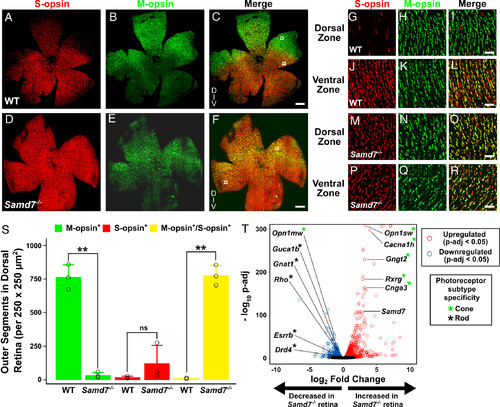
Dorsal M-cones are transformed into hybrid M/S-cones in the mouse Samd7−/− retina. (A–F) Flat mounts of adult WT and Samd7−/− mouse retina stained with S- and M-opsin antibodies demonstrate that S-opsin signal is increased throughout the dorsal region of Samd7−/− retina. (G–R) Close-up views of boxed insets from panels C and F reveal that S-opsin is up-regulated specifically in dorsal Samd7−/− M-opsin+ cones (i.e., M-cones are transformed into hybrid M/S-opsin+ cones). (S) The number of exclusively M-opsin+ cones in the dorsal Samd7−/− retina is reduced to a similar degree as the increase in the number of mixed M/S-opsin+ cones, supporting the conclusion that M-cones are transformed into hybrid M/S-opsin+ cones (mean ± SD; n = 3/genotype). (T) RNA-seq analysis of WT and Samd7−/− mouse retina demonstrates a reduction in rod-specific gene expression and an increase in cone-specific gene expression, with the exception of M-opsin (Opn1mw), which is somewhat reduced. Labeled genes were previously found to be dysregulated by qPCR analysis in another Samd7 mutant (38). Genes touching the top edge of the Volcano plot (e.g., Opn1sw) have p-adjusted values equal to zero (n = 3 retinas/genotype). Samd7minus19A mutants were used for flat-mount imaging and quantification in panels A–S. Samd7minus10A mutants were used for RNA-seq in panel T. [Scale bar in (A–F), 500 µm; Scale bar in (G–R), 20 µm.] D, V: Dorsal, Ventral. Statistical comparisons in S were performed using two-tailed, unpaired t test assuming unequal variance. **P < 0.005. SD, standard deviation. |
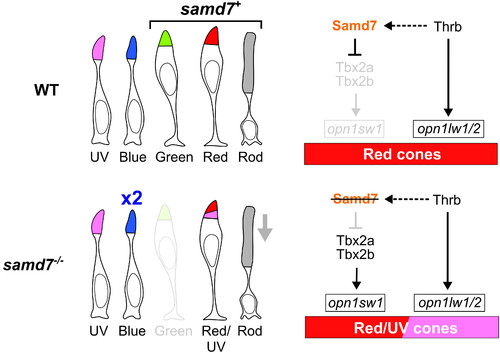
The role of samd7 in zebrafish photoreceptor specification. Diagram of cone subtypes in the WT and samd7−/− zebrafish retina. In the samd7−/− retina, red cones are transformed into hybrid red/UV-sensitive cones, green cones are absent, blue cones are approximately doubled, and the number of rods is greatly reduced. We propose that Samd7 represses UV-cone-specific gene expression by repressing tbx2a and tbx2b, two key regulators of opn1sw1 expression (4, 11, 12) Activation is indicated with an arrowhead, and repression with a flat bar. Black coloring indicates that the factor is expressed, and gray indicates that it is not. A dashed line represents a proposed regulatory relationship and a solid line represents a previously verified regulatory function. |