- Title
-
Gypenosides Alleviate Cone Cell Death in a Zebrafish Model of Retinitis Pigmentosa
- Authors
- Li, X., Alhasani, R.H., Cao, Y., Zhou, X., He, Z., Zeng, Z., Strang, N., Shu, X.
- Source
- Full text @ Antioxidants (Basel)
|
(A) Hematoxylin and eosin-stained image of retinal sections of wildtype (WT), rpgrip1 mutant and Gyp-treated rpgrip1 mutant zebrafish at 6 mpf. (B) Thickness of photoreceptor layer of the three zebrafish groups. Statistical comparisons between individual groups were carried out using one-way ANOVA followed by Bonferroni’s test. Data are displayed as mean ± SEM (n = 5 animals of each group). **** p < 0.0001. INL, inner nuclear layer; GCL, ganglion cell layer; ONL, outer nuclear layer; RPE, retinal pigment epithelial cells. PHENOTYPE:
|
|
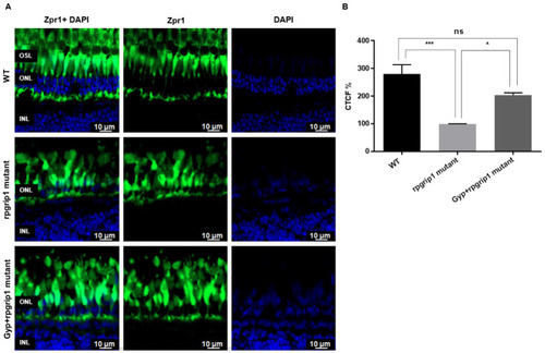
The effect of Gyp treatment on cone degeneration. (A) Immunostaining of retinal sections of wildtype, rpgrip1 mutant and Gyp-treated rpgrip1 mutant zebrafish at 6 mpf using zpr-1 antibody where zpr1 stained the cones in green and DAPI labelled nuclei in blue. (B) Intensity fluorescence signals (the corrected total cell fluorescence, CTCF) were measured using ImageJ. Statistical comparisons between individual groups were carried out using one-way ANOVA followed by Bonferroni’s test. Data are displayed as mean ± SEM (n = 5 animals of each group). Ns, no significance; * p < 0.05, *** p < 0.001. INL, inner nuclear layer; ONL, outer nuclear layer. |
|
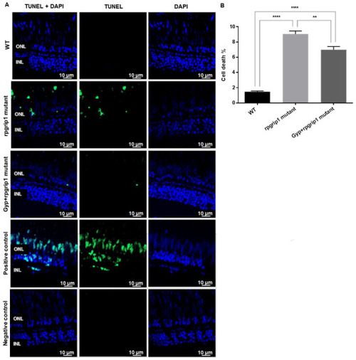
Significant decreases were found in cone apoptosis in rpgrip1 mutant zebrafish treated with Gyp. (A) Retinal sections of wildtype (WT), untreated (UT) rpgrip1 mutant and Gyp-treated rpgrip1 mutant zebrafish at 6 mpf were stained with TUNEL reagents. Nuclei of apoptotic cone cells were stained in green. DAPI labelled nuclei were stained in blue. (B) Quantification of apoptotic cells in above retinal sections were compared between the wildtype, rpgrip1 mutant and Gyp-treated rpgrip1 mutant zebrafish. Statistical comparisons between individual groups were carried out using one-way ANOVA followed by Bonferroni’s test. Data are displayed as mean ± SEM (n = 6 animals of each group). ** p < 0.01, **** p < 0.0001. INL, inner nuclear layer; ONL, outer nuclear layer. PHENOTYPE:
|
|
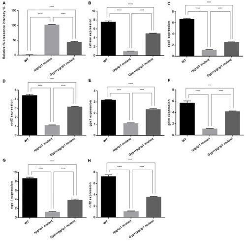
Effects of Gyp on antioxidant capacity in rpgrip1 mutant zebrafish eyes. (A) ROS generation in the eyes of wildtype (WT), untreated (UT) and Gyp-treated rpgrip1 mutant zebrafish at 6 mpf. (B–H) Expression of catalase, sod1, sod2, gpx1, gclm, nqo-1, gclm and nrf2 was determined using qRT-PCR. Statistical comparisons between individual groups were carried out using one-way ANOVA followed by Bonferroni’s test. Data are displayed as mean ± SEM (n = 6 animals of each group). ** p < 0.01, **** p < 0.0001. |
|
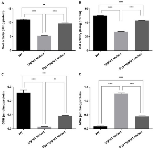
Sod (A) and catalase (B) activities, and GSH level (C) were markedly increased but MDA level (D) was significantly decreased in Gyp-treated rpgrip1 mutant zebrafish eye samples. WT, wildtype zebrafish; UT, untreated rpgrip1 mutant zebrafish. Statistical comparisons between individual groups were carried out using one-way ANOVA followed by Bonferroni’s test. Data are displayed as mean ± SEM (n = 6 animals of each group). ** p < 0.01, *** p < 0.001, **** p < 0.0001. PHENOTYPE:
|
|
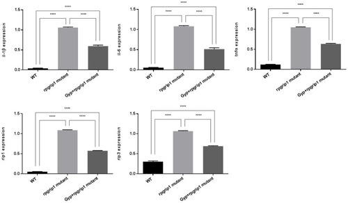
Gyp treatment reduced the expression of inflammation associated genes in rpgrip1 mutant zebrafish eye samples. Expression of il-1β, il-6, tnfα, rip1 and rip3 in eyes of wildtype (WT), untreated (UT) and Gyp-treated rpgrip1 mutant zebrafish at 6 mpf was determined by qRT-PCR. Statistical comparisons between individual groups were carried out using one-way ANOVA followed by Bonferroni’s test. Data are displayed as mean ± SEM (n = 6 animals of each group). **** p < 0.0001. |
|
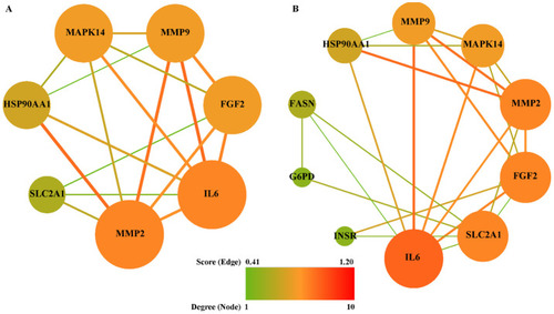
PPI network between common targets. (A) Inflammation, (B) oxidative stress. The nodes represented targets and edges represented interaction among targets. The node size and color were represented with degree, while edge size and color were represented with combined score. |


