
IMAGE
Figure 1
- ID
- ZDB-IMAGE-210726-51
- Publication
- Li et al., 2021 - Gypenosides Alleviate Cone Cell Death in a Zebrafish Model of Retinitis Pigmentosa
- All Figures
- Figures for Li et al., 2021
Image
|
Figure Caption
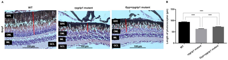
Figure 1 (A) Hematoxylin and eosin-stained image of retinal sections of wildtype (WT), rpgrip1 mutant and Gyp-treated rpgrip1 mutant zebrafish at 6 mpf. (B) Thickness of photoreceptor layer of the three zebrafish groups. Statistical comparisons between individual groups were carried out using one-way ANOVA followed by Bonferroni’s test. Data are displayed as mean ± SEM (n = 5 animals of each group). **** p < 0.0001. INL, inner nuclear layer; GCL, ganglion cell layer; ONL, outer nuclear layer; RPE, retinal pigment epithelial cells.
Figure Data
Acknowledgments
This image is the copyrighted work of the attributed author or publisher, and
ZFIN has permission only to display this image to its users.
Additional permissions should be obtained from the applicable author or publisher of the image.
Full text @ Antioxidants (Basel)