FIGURE SUMMARY
Title

The stem-like STAT3-responsive cells of zebrafish intestine are WNT/β-catenin dependent

Authors
Peron, M., Dinarello, A., Meneghetti, G., Martorano, L., Facchinello, N., Vettori, A., Licciardello, G., Tiso, N., Argenton, F.
Source
Full text @ Development

Generation of Tg(7xStat3-Hsv.Ul23:EGFP) fish and characterization of their expression pattern. (A) Scheme of the Tol-2 vectors used to generate the Tg(7xStat3-Hsv.Ul23:EGFP) reporter. (B,B′) Diffused EGFP is detectable in early stage embryos obtained by outcrossing transgenic females (B), not with transgenic males (B′). (C-F) At 22 hpf, EGFP expression is detectable in the anterior telencephalic region (t), the primordial midbrain hindbrain boundary (mhb), the hindbrain (h), the primitive neuromasts (n) and in the haematopoietic tissue (ht). (G,H) At 48 hpf, EGFP expression is mostly located in the optic tectum (TeO) and in the hindbrain (h). (I,J) Starting from 4 dpf, EGFP expression is detectable in the developing intestine in isolated pear-shaped cells (I); the intestinal fluorescence lasts throughout adulthood (J).

Tg(7xStat3:EGFP) is a bona fide Stat3 pathway reporter. (A) Whole mount in situ hybridization detection of EGFP mRNA in the optic tectum of 48 hpf embryos treated with inhibitors of the Stat3 pathway, AG-490 and LLL12; control embryos were treated with DMSO. (A′) Percentage of samples displaying EGFP mRNA expression in the optic tectum (three independent biological samples; n=40). (B) Fluorescent image of Tg(7xStat3:EGFP) intestine at 6 dpf after 3 days of either 60 µM AG-490 or DMSO treatment. (B′) EGFP fluorescence quantification in the intestine of AG-490 and control larvae after 3 days of 60 µM AG-490 administration (n=20). (C) qRT-PCR analysis of stat3, socs3a and EGFP expression in EGFP-positive and EGFP-negative cells taken from adult intestines. (D) Immunofluorescence against mouse Stat3 (red spots) and colocalization with EGFP fluorescence of Tg(7xStat3:EGFP) larvae injected with CMV-mStat3C plasmid. (E) Live image of the intestine of 7 dpf stat3−/−/Tg(7xStat3:EGFP) and stat3+/+/Tg(7xStat3:EGFP) siblings. (E′) EGFP fluorescence quantification in the intestine of wild-type (WT) and stat3−/− Tg(7xStat3:EGFP) larvae at 7 dpf (n=12, P=0.0211). (E″) qRT-PCR analysis of EGFP expression in 7 dpf stat3−/−/Tg(7xStat3:EGFP) larvae with respect to the stat3+/+/Tg(7xStat3:EGFP) sibling (three biological samples). All statistical analyses were performed by unpaired t-test; *P<0.05, **P<0.01, ***P<0.001, ****P<0.0001. Graphs indicate mean±s.e.m. Scale bars: 100 μm (A,B,E), 10 μm (D).

Stat3 pathway is active in proliferating cells of zebrafish haematopoietic tissue and optic tectum. (A-A″) Fluorescence co-localization (A″) using α-EGFP Ab (A) and EdU proliferation assay (A′) in the haematopoietic tissue (dashed line) of 22 hpf Tg(7xStat3:EGFP) reporter embryos. (B-B″) Fluorescence co-localization (B″) between fish using α-EGFP probe (B) and EdU proliferation assay (B′) in the optic tectum (TeO; dashed line) of 48 hpf embryos. (C,C′) In vivo fluorescence of Tg(7xStat3:EGFP) reporter activity in the TeO of embryos treated with LY364947 inhibitor between 24 and 48 hpf (C′) compared with DMSO-treated controls (C). (C″) Relative fluorescence intensity in the TeO of 48 hpf Tg(7xStat3:EGFP) embryos described in C (n=15, P=0.0014). (D,D′) Whole mount in situ hybridization detection of pcna mRNA in the TeO of 48 hpf embryos treated with LY364947 inhibitor between 24 and 48 hpf (D′) compared with DMSO-treated controls (D). (D″) pcna mRNA expression in the embryos described in D (P=0.038). All statistical analyses were performed by unpaired t-test; *P<0.05, **P<0.01. Graphs indicate mean±s.e.m. Scale bar: 100 μM.

Stat3 pathway is active in rapidly proliferating intestinal cells during zebrafish larval development. (A-A″) Co-localization (A″) using α-EGFP Ab (A) and EdU proliferation assay (A′) in the developing intestine (dashed line) of 5 dpf Tg(7xStat3:EGFP) larvae. (B,B′) Label retention assay using double transgenic Tg(7xStat3:EGFP)/Tg(HSP70:H2B:mRFP) 5 dpf larvae. The ubiquitous RFP label, accumulated in 100% of intestinal cells at 6 hpHS, is completely lost in EGFP-expressing cells at 48 hpHS, where no EGFP/RFP co-localization is appreciable (B′). (B″) RFP loss by Tg(7xStat3:EGFP)-positive intestinal cells. Three independent biological replicates were performed.

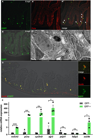
Stat3 is active in intestinal FBC cells of adult zebrafish. (A-A″) Co-localization (A″) using α-EGFP Ab (green) (A) and α-PCNA Ab (red) (A′) staining of adult Tg(7xStat3:EGFP) intestine. (B) Staining with α-EGFP Ab on a transversal section of adult Tg(7xStat3:EGFP) intestine, showing that all Stat3-positive cells are located at the base of the intervillus pocket. (C,C′) Immunogold staining with α-EGFP Ab on adult Tg(7xStat3:EGFP) intestinal sections observed by TEM. A gold-labelled cell is surrounded with a white striped line (C). High magnification of gold-labelled cytoplasm belonging to triangular-shaped fold base columnar (FBC) cell; inset is zoom of boxed area, showing gold dots (black arrowheads). (D) Staining with α-EGFP Ab (green) and α-Sox9b Ab (red) of adult Tg(7xStat3:EGFP) intestine. (E) qRT-PCR analysis of notch2, pcna, cylcinD1, agr2, pept1, fabp2 and sox9b expression in EGFP-positive and EGFP-negative cells taken from adult intestines. Statistical analysis was performed by unpaired t-test; *P<0.05, ***P<0.001, ****P<0.0001; ns, not significant. Error bars indicate s.e.m.

EXPRESSION / LABELING:
Genes:
Antibody:
Fish:
Anatomical Term:
Stage: Adult

Stat3 is required for intestinal homeostasis and larvae survival during development. (A) Percentage of the different genotypes found at different time points in stat3+/− incross; stat3−/− mutants were never found after 25 dpf, with the exception of a single animal that died at 51 dpf (shown in B). Statistical analysis was performed by chi-square test to compare proportions. (B) Haematoxylin-eosin staining of longitudinal sections at different larval stages (indicated on the left); arrowhead indicates the intestinal epithelium. Compared with normal fish, mutants display flattening of the intestinal epithelium. (C) Percentage of individuals at different stages presenting either normal intestine or degenerated intestine lacking intestinal folds. *P<0.05; **P<0.01.

Tcf7l2 (Tcf4) is required for development of Stat3-responsive cells of zebrafish larvae intestine and the Stat3 pathway is activated ectopically in intestinal adenomas of apchu745 mutants. (A,A′) In vivo EGFP fluorescence in the intestine of 6 dpf Tg(7xStat3:EGFP)/tcf7l2hu892/hu892, Tg(7xStat3:EGFP)/tcf7l2+/hu892 and Tg(7xStat3:EGFP)/tcf7l2+/+ siblings (A) and measurement of integrated density (A′) (n=16). (B) qPCR analysis of il6, gp130, jak2a, jak2b and stat3 mRNA expression from tcf7l2+/+ and tcf7l2hu892/hu892 sibling larvae. (C,C′) Effect of XAV treatment on Tg(7xStat3:EGFP) embryos from 48 to 78 hpf: measurement of the integrated density of the fluorescence (C) and measurement of the number of GFP+ cells (C′). (D,E) Haematoxylin-eosin staining on paraffin embedded transversal section of zebrafish apchu745 intestine at 12 months post fertilization, displaying both normal tissue (n) and hyperplastic adenomas (a). (D′,D″) Staining of a sequential intestinal section of D using α-EGFP (green) (D′) and α-PCNA (red) (D″) Abs. (E′,E″) Double staining using α-EGFP Ab (green) and α-PCNA Ab (red) of a sequential intestinal section of E. All statistical analyses were performed by unpaired t-test. *P<0.05, ***P<0.001; ns, not significant. Error bars indicate s.e.m.

ZFIN is incorporating published figure images and captions as part of an ongoing project. Figures from some publications have not yet been curated, or are not available for display because of copyright restrictions.

EXPRESSION / LABELING:
Genes:
Fish:
Anatomical Term:
Stage: Day 6
PHENOTYPE:
Fish:
Observed In:
Stage: Day 6

ZFIN is incorporating published figure images and captions as part of an ongoing project. Figures from some publications have not yet been curated, or are not available for display because of copyright restrictions.

PHENOTYPE:
Fish:
Observed In:
Stage: Adult
Acknowledgments
This image is the copyrighted work of the attributed author or publisher, and ZFIN has permission only to display this image to its users. Additional permissions should be obtained from the applicable author or publisher of the image. Full text @ Development