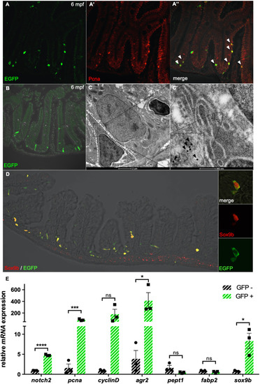
Stat3 is active in intestinal FBC cells of adult zebrafish. (A-A″) Co-localization (A″) using α-EGFP Ab (green) (A) and α-PCNA Ab (red) (A′) staining of adult Tg(7xStat3:EGFP) intestine. (B) Staining with α-EGFP Ab on a transversal section of adult Tg(7xStat3:EGFP) intestine, showing that all Stat3-positive cells are located at the base of the intervillus pocket. (C,C′) Immunogold staining with α-EGFP Ab on adult Tg(7xStat3:EGFP) intestinal sections observed by TEM. A gold-labelled cell is surrounded with a white striped line (C). High magnification of gold-labelled cytoplasm belonging to triangular-shaped fold base columnar (FBC) cell; inset is zoom of boxed area, showing gold dots (black arrowheads). (D) Staining with α-EGFP Ab (green) and α-Sox9b Ab (red) of adult Tg(7xStat3:EGFP) intestine. (E) qRT-PCR analysis of notch2, pcna, cylcinD1, agr2, pept1, fabp2 and sox9b expression in EGFP-positive and EGFP-negative cells taken from adult intestines. Statistical analysis was performed by unpaired t-test; *P<0.05, ***P<0.001, ****P<0.0001; ns, not significant. Error bars indicate s.e.m.
|
