- Title
-
DEAD-Box Protein Ddx46 Is Required for the Development of the Digestive Organs and Brain in Zebrafish
- Authors
- Hozumi, S., Hirabayashi, R., Yoshizawa, A., Ogata, M., Ishitani, T., Tsutsumi, M., Kuroiwa, A., Itoh, M., and Kikuchi, Y.
- Source
- Full text @ PLoS One
|
Phenotype of the morha4 mutant. (A–F) Lateral (A–D) and dorsal (E, F) views of live WT and morha4 larvae at 5.5 dpf. The swim bladder failed to inflate (arrows in A, B), the intestine lacked folds (arrowheads in C, D), and the retinae were reduced in size (brackets in E, F) in the morha4 mutant. Conversely, somite formation in the morha4 mutant appeared normal (arrowheads in A, B). (G–L) Sagittal sections of 5.5-dpf larvae stained with hematoxylin and eosin. The intestine lacked folds and was thin walled (arrowheads in G, H), and the exocrine pancreas (blue dotted lines in I, J) and liver (blue dotted lines in K, L) were small in the morha4 mutant. In contrast, the endocrine pancreas (blue dotted lines in I, J) in WT larvae was indistinguishable from that in morha4 larvae. Scale bars, 50 μm. (M–P) Dorsal views, anterior to the top (M, N). Lateral views, anterior to the left (O, P). Apoptotic cells were detected using the TUNEL method. An increase in apoptotic cells was evident in the brain, retinae, and posterior intestine of the morha4 larvae (white arrowheads in O, P) compared to WT larvae, but not in the morha4 somite (white arrows in O, P). en, endocrine pancreas; ex, exocrine pancreas. PHENOTYPE:
|
|
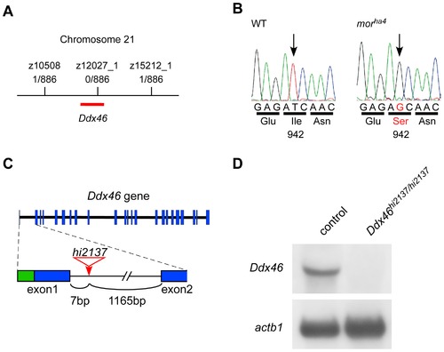
Identification of the mor gene and analysis of the hi2137 allele. (A) Meiotic and physical map schematic of the mor locus on chromosome 21. The number of recombinants and larvae genotyped is shown for each microsatellite marker. (B) Sequencing cDNA from WT and morha4 larvae revealed a nucleotide exchange from T to G, which resulted in an Ile-to-Ser transition at amino acid 942 in the morha4 mutant. (C) Genomic structure of the Ddx46 gene showing the viral insertion site in the hi2137 allele (red). Exons are boxes, with coding and non-coding sequences in blue and green, respectively. The viral insertion (red arrow) occurs in the first intron between exons 1 and 2. (D) Northern blot analysis of Ddx46hi2137/hi2137 mutants and control larvae at 3.5 dpf. No Ddx46 transcript was found in the Ddx46hi2137/hi2137 mutants, whereas the level of actb1 transcript in the mutants was the same as that in control larvae. Control larvae were sibling WT or Ddx46hi2137/+ larvae and had normal phenotypes. EXPRESSION / LABELING:
|
|
Defects of exocrine pancreas formation in both morha4/ha4 and Ddx46hi2137/hi2137 mutants are rescued by the overexpression of Ddx46 mRNA but not mutated Ddx46 mRNA. (A) Scheme of the Ddx46 protein structure. The yellow, red, and orange boxes indicate the N-terminal, DEAD-box helicase, and C-terminal domain, respectively. Mutations were introduced into the Ddx46 protein; in Ddx46-I942S, an isoleucine in the C-terminal domain of Ddx46 was changed to serine, which is the same mutation as that in the morha4 mutant; in Ddx46-K402A, GKT in motif I, which is important for ATPase activity in Ddx46 homologues, was changed to GAT. (B–I) All dorsal views, anterior to the top. The expression of try, a molecular marker for the exocrine pancreas, was examined using whole-mount in situ hybridization at 3.5 dpf. The try expression in the exocrine pancreas was markedly reduced in egfp mRNA-injected morha4/ha4 (C) and Ddx46hi2137/hi2137 mutants (F) compared to egfp mRNA-injected control larvae (B, E). The try expression was rescued in the Ddx46 mRNA-injected morha4/ha4 (D) and Ddx46hi2137/hi2137 mutants (G), whereas no rescue was achieved by the overexpression of Ddx46-I942S (H) or Ddx46-K402A (I) mRNA into Ddx46hi2137/hi2137 mutants. Control larvae-sibling WT or morha4/+ larvae (B–D), sibling WT or Ddx46hi2137/+ larvae (E–I)-had normal phenotypes. EXPRESSION / LABELING:
PHENOTYPE:
|
|
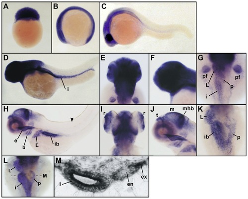
Ddx46 expression in the developing zebrafish. (A–K) Ddx46 expression was examined using whole-mount in situ hybridization in WT embryos or larvae at the 128-cell (A), 6-somite (B), 1-dpf (C), 2-dpf (D–G), and 4-dpf (H–K) stages. Lateral view, animal pole to the top (A). Lateral views, anterior to the left (B, C, D, F, H, J). Dorsal views, anterior to the top (E, G, I, K). The Ddx46 transcript was maternally supplied and continued to be expressed ubiquitously during the somitogenesis stages (A, B). By 1 dpf, Ddx46 expression became restricted to the head region (C). At 2 dpf, strong Ddx46 expression was prominent in the head, pectoral fin bud, and digestive organs (D–G). At 4 dpf, Ddx46 expression was further restricted to the retina, telencephalon, midbrain, midbrain-hindbrain boundary, branchial arch, esophagus, liver, pancreas, and intestinal bulb (H–K). No Ddx46 transcript was detected in the somite (arrowhead in H). (L, M) Ddx46 expression was examined using whole-mount in situ hybridization in WT larvae at 3 dpf. Dorsal views, anterior to the top (L). A transverse section was cut at the level indicated by the black dotted line in L. The section revealed Ddx46 expression in the intestine and exocrine pancreas, but not in the endocrine pancreas (M). b, branchial arches; e, esophagus; en, endocrine pancreas; ex, exocrine pancreas; i, intestine; ib, intestinal bulb; L, liver; m, midbrain; mhb, midbrain-hindbrain boundary; p, pancreas; pf, pectoral fin bud; r, retina; t, telencephalon. |
|
Expression of molecular markers for digestive organs and brain is reduced in the Ddx46hi2137/hi2137 mutant. (A–D) The expression of dla and her6 was examined using whole-mount in situ hybridization at 3 dpf. All lateral views, anterior to the left. (E–L) The expression of fabp2, fabp10a, ptf1a, and ins was examined using whole-mount in situ hybridization at 3.5 dpf. All dorsal views, anterior to the top. In the Ddx46hi2137/hi2137 mutants, the intensity and area of dla, her6, fabp2, fabp10a, and ptf1a expression were markedly reduced at 3 or 3.5 dpf (A–J; arrowheads in H, J). In contrast, the ins expression in the Ddx46hi2137/hi2137 mutant did not change at these developmental stages (K, L). (M–P) Transverse sections of 3.5-dpf Ddx46hi2137/hi2137 mutant larvae stained with hematoxylin and eosin. The transverse sections were cut at the levels indicated by black dotted lines in E–L. The tissues in the intestinal bulb, liver, and exocrine pancreas were still present in the Ddx46hi2137/hi2137 mutant larvae at 3.5 dpf. Scale bars, 50 μm. en, endocrine pancreas; ex, exocrine pancreas; ib, intestinal bulb; L, liver. Control larvae were sibling WT or Ddx46hi2137/+ larvae and had normal phenotypes. EXPRESSION / LABELING:
PHENOTYPE:
|
|
Ddx46 deficiency affects pre-mRNA splicing in the digestive organs and brain. (A–H) Scheme of the dla, her6, ptf1a, and fabp10a pre-mRNA regions analyzed for splicing (boxes, exons; lines, introns; arrows, primers) (A, C, E, G). The splicing status of dla, her6, ptf1a, and fabp10a pre-mRNA was monitored using RT-PCR with the primers indicated in scheme A, C, E, and G, respectively. Unspliced dla, her6, ptf1a, and fabp10a mRNAs were retained in the Ddx46hi2137/hi2137 mutant (mut) larvae compared to the control (con) larvae (arrowheads in B, D, F, H). Unspliced and spliced PCR products were verified by sequencing. +RT refers to the validation reaction itself, and RT represents the respective control reaction without reverse transcriptase. actb1 is a loading control by using primers designed in the exon 6. M, DNA size markers (sizes in bp); the asterisks point to nonspecific PCR products. Control larvae were sibling WT or Ddx46hi2137/+ larvae and had normal phenotypes. PHENOTYPE:
|
|
The size of the exocrine pancreas is reduced in the morha4 mutant. (A, B) High-power, lateral views of the immunostained exocrine pancreas from 5.5 dpf WT and morha4 larvae. Both larvae were processed for carboxypeptidase A immunohistochemistry. The size of the exocrine pancreas was markedly reduced in the morha4 mutant compared to the WT larva. Scale bars, 50 μm. |
|
Transheterozygote (morha4/Ddx46hi2137) of morha4 and Ddx46hi2137 shows the phenocopy of the morha4 mutant. (A–F) Lateral (A–D) and dorsal (E, F) views of live control and morha4/Ddx46hi2137 larvae at 5 dpf. The swim bladder failed to inflate (arrows in A, B), the intestine lacked folds (arrowheads in C, D), and the retinae were reduced in size (brackets in E, F) in the morha4/Ddx46hi2137 mutant. Conversely, somite formation in the morha4/Ddx46hi2137 mutant appeared normal (arrowheads in A, B). Control larvae were sibling WT, morha4/+ or Ddx46hi2137/+ larvae and had normal phenotypes. PHENOTYPE:
|
|
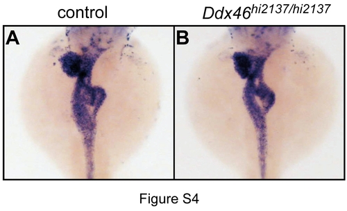
Expression of foxa3 is unaffected in the Ddx46hi2137/hi2137 mutant at 2.5 dpf. (A, B) Expression of foxa3 was examined using whole-mount in situ hybridization. Dorsal views, anterior to the top. The foxa3 expression in control larvae (A) was indistinguishable from that in the Ddx46hi2137/hi2137 mutant (B) at 2.5 dpf. Control larvae were sibling WT or Ddx46hi2137/+ larvae and had normal phenotypes. EXPRESSION / LABELING:
|
|
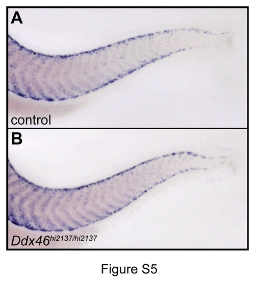
Expression of myod1 is normal in the Ddx46hi2137/hi2137 mutant. (A, B) Expression of myod1 was examined using whole-mount in situ hybridization. Lateral views, anterior to the left. The myod1 expression in control larvae (A) was indistinguishable from that in the Ddx46hi2137/hi2137 mutant (B) at 3.5 dpf. Control larvae were sibling WT or Ddx46hi2137/+ larvae and had normal phenotypes. EXPRESSION / LABELING:
|
|
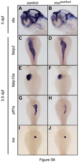
Expression of molecular markers for digestive organs and brain is reduced in the morha4/ha4 mutant. (A–B) The expression of dla was examined using whole-mount in situ hybridization at 3 dpf. All lateral views, anterior to the left. (C–J) The expression of fabp2, fabp10a, ptf1a, and ins was examined using whole-mount in situ hybridization at 3.5 dpf. All dorsal views, anterior to the top. Although the expression of dla, fabp2, and fabp10a was slightly reduced, the ptf1a expression was markdly reduced at 3 or 3.5 dpf in the morha4/ha4 mutants (A–H). In contrast, the ins expression in the morha4/ha4 mutant did not change at these developmental stages (I, J). Control larvae were sibling WT or morha4/+ larvae and had normal phenotypes. EXPRESSION / LABELING:
PHENOTYPE:
|
|
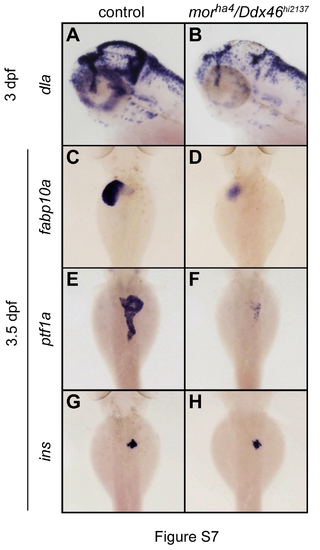
Expression of molecular markers for digestive organs and brain is also reduced in the transheterozygote morha4/Ddx46hi2137 mutant. (A, B) The expression of dla was examined using whole-mount in situ hybridization at 3 dpf. All lateral views, anterior to the left. (C–H) The expression of fabp10a, ptf1a, and ins was examined by whole-mount in situ hybridization at 3.5 dpf. All dorsal views, anterior to the top. The intensity and area of dla, fabp10a, and ptf1a expression were markedly reduced at 3 or 3.5 dpf in the morha4/Ddx46hi2137 mutants. In contrast, ins expression in this transheterozygote was unchanged at these developmental stages. These phenotypes are the same as those of the Ddx46hi2137/hi2137 mutant. Control larvae were sibling WT, morha4/+, or Ddx46hi2137/+ larvae and had normal phenotypes. EXPRESSION / LABELING:
PHENOTYPE:
|
|
Expression of various molecular markers for digestive organs and brain is reduced in the Ddx46hi2137/hi2137 mutant. (A–F) The expression of her4, neurog1, and neurod for brain was examined using whole-mount in situ hybridization at 3 dpf. All lateral views, anterior to the left. (G–N) The expression of hlxb9la, cpa5, gata6, and dhrs9 for digestive organs was examined using whole-mount in situ hybridization at 3.5 dpf. All dorsal views, anterior to the top. In the Ddx46hi2137/hi2137 mutants, the intensity and area of all of these gene expressions were markedly reduced at 3 or 3.5 dpf. Control larvae were sibling WT or Ddx46hi2137/+ larvae and had normal phenotypes. EXPRESSION / LABELING:
|
|
Pre-mRNA splicing of the housekeeping gene actb1, but not b2m, is unaffected in the Ddx46hi2137/hi2137 mutant. (A–D) Scheme of the b2m and actb1 pre-mRNA regions analyzed for splicing (boxes, exons; lines, introns; arrows, primers) (A, C). The splicing status of b2m and actb1 pre-mRNA was monitored using RT-PCR with the primers indicated in scheme A and C, respectively. Total RNA was isolated from the heads of Ddx46hi2137/hi2137 mutants (mut) and control (con) larvae. Unspliced b2m mRNAs were retained in the Ddx46hi2137/hi2137 mutants compared to the control larvae (arrowheads in B), whereas the splicing of actb1 was unaffected in the Ddx46hi2137/hi2137 mutants (arrowheads in D). Unspliced and spliced PCR products were verified by sequencing. +RT refers to the validation reaction itself, and -RT represents the respective control reaction without reverse transcriptase. 18S rRNA was used as a loading control. M, DNA size markers (sizes in bp). Control larvae were sibling WT or Ddx46hi2137/+ larvae and had normal phenotypes. PHENOTYPE:
|


