
- Title
-
Integrity of the midbrain region is required to maintain the diencephalic-mesencephalic boundary in zebrafish no isthmus/pax2.1 mutants
- Authors
- Scholpp, S. and Brand, M.
- Source
- Full text @ Dev. Dyn.
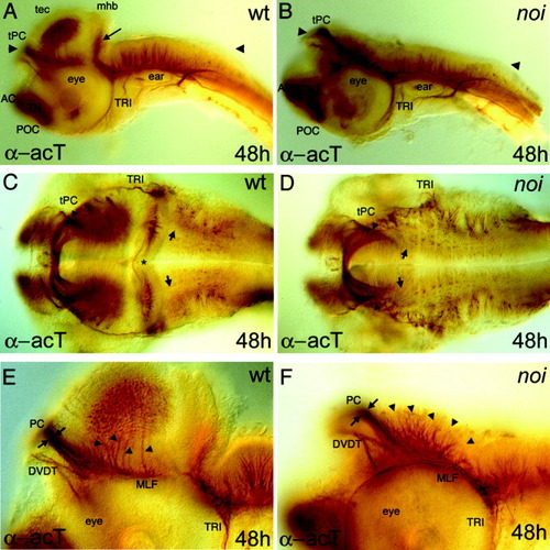
The posterior commissure (PC) and the medial longitudinal fasciculus (MLF) are present but modified in noi mutant embryos. Immunochemical labelling of acetylated tubulin was used to study axon formation in 48 hours (48h) postfertilization embryos, oriented anterior to the left. A,B: A lateral view of whole-mounted embryos. The tectum and the cerebellar fold, including their neurons, e.g., the nervus trochlearis, are absent in the mutant. Arrowheads in A and B mark the layer of the dorsal section in C and D, respectively. C,D: The PC, which demarcates the diencephalic/mesencephalic boundary, is still present. The PC looks more condensed in the mediolateral direction (arrowheads) and hindbrain neurons, visualized in small dots expand anteriorly (arrows). E,F: Higher magnification of the lateral view of the mutant embryo shows additional axons emerging from the dorsal midbrain area (arrowheads) and joining the medial longitudinal fasciculus (MLF). In the anteroposterior direction, the PC appears larger and defasciculated (E,F; arrows). AC, anterior commissure; DVDT, dorsoventral diencephalic tract; mhb, midbrain-hindbrain boundary; tPC, tract of the posterior commissure; POC, postoptic commissure; tec, tectum opticum; TN, telencephalic nucleus; TRI, trigeminal nerve, tro, trochlear nerve; wt, wild-type. PHENOTYPE:
|
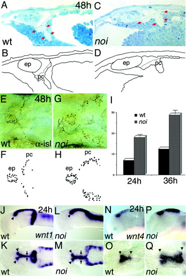
The territory of the dorsal forebrain and midbrain is altered in noi mutant embryos. Histologic, immunochemical, and in situ hybridisation (ISH) analysis was used to study the dorsal forebrain and midbrain in noi mutant embryos. The embryos were oriented with anterior to the left. A,B: A parasagittal section through the dorsal part of the forebrain-midbrain region of a 48 hours postfertilization (hpf; 48h) wild-type (wt) zebrafish embryo shows the location of the epiphysis (ep) and the posterior commissure (pc). C,D: In the mutant, the ep and the pc seem enlarged in the anteroposterior direction marked with red arrows in C. E-H: A dorsal view of the nucleus of the posterior commissure interneurons stained with an α-islet (αisl) antibody indicates that the number of neurons is increased and the territory is expanded posteriorly. I: Already at 24 hpf and at 36 hpf, a significantly increased number of neurons is observed between wild-type siblings and in noi mutant embryos. J-M: A lateral and dorsal view of an ISH of wnt1 shows a fusion of the midbrain and hindbrain pattern and the missing expression in the ventral part of the midbrain-hindbrain boundary. N-Q: The expression wnt4 shows an expansion of these dorsal diencephalic markers into the midbrain territory (arrowheads). PHENOTYPE:
|
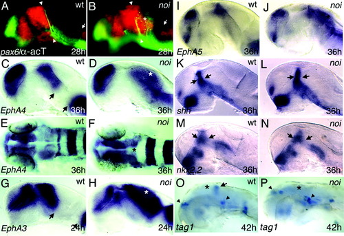
Marker analysis in wild-type (wt) and noi mutant embryos suggests a respecification of midbrain tissue. In situ hybridisation with the indicated marker genes at the given stages (h, hours postfertilisation), lateral views (except dorsal views in E, F) with anterior to the left; asterisks mark the position of the misspecified midbrain. A: Staining for pax6.1, a marker for the forebrain and basal hindbrain, combined with an acetylated tubulin staining to visualize outgrowing axons. The posterior commissure (PC, marked by white arrowheads) is located at the diencephalic-mesencephalic boundary in the pax6.1+ domain. B: In the noi mutant, pax6.1 expression is observed in the territory of the presumptive midbrain and new branches of the PC (yellow arrowheads) are visible more posterior to the endogenous position. Similar to the forebrain, the hindbrain expression domain expands anteriorly into the midbrain territory (A,B, white arrows). Markers of the Ephrin family show a similar phenomenon: EphA4 (C,E), EphA3 (G), and EphA5 (I) respect the boundary between the diencephalon and the mesencephalon (black arrows). D,F,H,J: In the noi mutant, the expression expands into the misspecified midbrain area. K-N: Marker genes of the zona limitans intrathalamica, such as shh and nkx2.2, show no difference between noi mutant and wild-type siblings, suggesting a correct formation of this anterior region between the prosomeres 2 and 3 (arrows). O: tag1 marks a subset of developing neurons in the cerebellar anlage (arrow). P: This expression domain is missing in the noi mutants, consistent with the loss of cerebellar identity in the noi mutants. However, the expression domains in the dorsal forebrain and in the anterior hindbrain are not altered (indicated by arrowheads). |
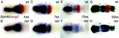
Time course of marker expansion in noi mutant embryos. Double in situ hybridisation of wild-type (wt) and noi mutants with EphA5 in blue and eng2 in red. A,B: At the four-somite stage (4ss), there is no difference in forebrain marker expression between wild-type and mutants. C,D: At 7ss, the first change of marker expression is visible: EphA5 expression starts in the anterior midbrain. eng2 is used to distinguish wild-type embryos and mutant embryos, because the expression of eng2 is absent in noi mutant embryos. E,F: At 12ss, EphA5 is clearly expanded into the midbrain. G,H: The situation at 20ss in which the whole respecified midbrain expresses EphA5. |
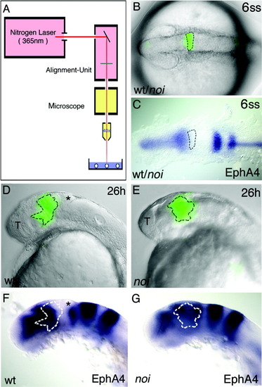
Presumptive midbrain tissue of noi mutant embryos is transformed into forebrain fate. Wild-type (wt) and noi mutant embryos were injected with caged fluorescein at the one-cell stage. A: At the six-somite stage (6ss), a nitrogen laser with a wavelength of 365 nm was used to activate the caged fluorescein in cells located at the position of the anterior midbrain primordium, as identified by comparison to the fate map and to gene expression data. B: A brightfield picture of an embryo at 6ss superimposed onto the picture of the uncaged, fluorescein-labelled cells in the midbrain primordium of the same embryo. C: A comparison with the expression pattern of EphA4 in another embryo at the same stage indicates that the uncaged cell clone is located mainly in the area of the EphA4-negative anterior midbrain. D: At 26 hours postfertilisation (h), the position of the progeny of this cell clone was identified and is shown again superimposed with the brightfield picture. F: The same embryo was used for in situ hybridisation analysis with EphA4. The cell clone covers the region of the posterior forebrain (EphA4-positive) and the anterior midbrain (EphA4-negative). E,G: The same procedure was done with noi mutant embryos. The cell clone in the noi mutant embryos exclusively expresses EphA4, suggesting a transformation of cell fate. T, telencephalon. |

Unillustrated author statements EXPRESSION / LABELING:
|