
|
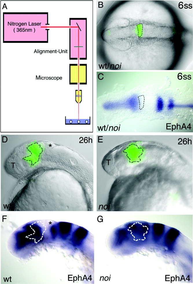
Fig. 5 Presumptive midbrain tissue of noi mutant embryos is transformed into forebrain fate. Wild-type (wt) and noi mutant embryos were injected with caged fluorescein at the one-cell stage. A: At the six-somite stage (6ss), a nitrogen laser with a wavelength of 365 nm was used to activate the caged fluorescein in cells located at the position of the anterior midbrain primordium, as identified by comparison to the fate map and to gene expression data. B: A brightfield picture of an embryo at 6ss superimposed onto the picture of the uncaged, fluorescein-labelled cells in the midbrain primordium of the same embryo. C: A comparison with the expression pattern of EphA4 in another embryo at the same stage indicates that the uncaged cell clone is located mainly in the area of the EphA4-negative anterior midbrain. D: At 26 hours postfertilisation (h), the position of the progeny of this cell clone was identified and is shown again superimposed with the brightfield picture. F: The same embryo was used for in situ hybridisation analysis with EphA4. The cell clone covers the region of the posterior forebrain (EphA4-positive) and the anterior midbrain (EphA4-negative). E,G: The same procedure was done with noi mutant embryos. The cell clone in the noi mutant embryos exclusively expresses EphA4, suggesting a transformation of cell fate. T, telencephalon.