
- Title
-
Intra-endodermal interactions are required for pancreatic beta cell induction
- Authors
- Chung, W.S., and Stainier, D.Y.
- Source
- Full text @ Dev. Cell
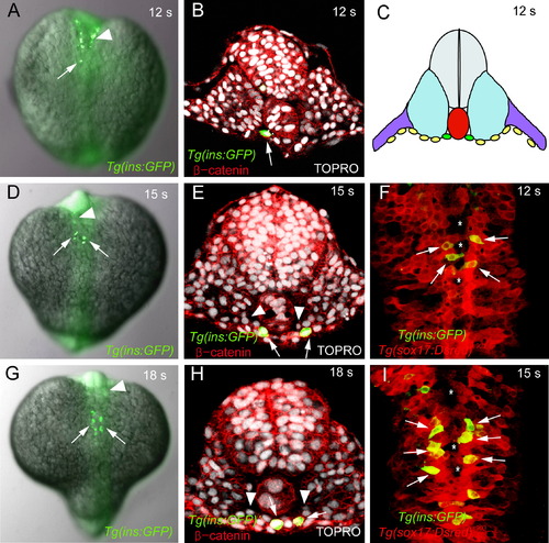
Initial Position of Tg(ins:GFP)-Expressing Cells 12- (A?C, and F), 15- (D?E, and I), and 18- (G?H) somite stage (corresponding to 15, 16.5, and 18 hpf, respectively) Tg(ins:GFP) embryos showing GFP (green; [A?B and D?I]), TOPRO (white; [B, E, and H]), β-catenin (red; [B, E, and H]) and Tg(sox17:DsRed)s903 (red; [F and I]) staining. Dorsal views of fixed embryos (A, D, and G), and confocal images of transverse sections (B, E, and H) and of ventral views (F and I). (A) One Tg(ins:GFP)-expressing cell (arrow) is present in the endoderm of this 12-somite stage embryo. Cells in the anterior neural tube (arrowhead) also express the ins:GFP transgene. (B) The Tg(ins:GFP)-expressing cell (arrow) is located in the medio-lateral axis between the notochord and left somite. (C) Schematic diagram of a transverse section showing Tg(ins:GFP)-expressing cells (green) within the endodermal sheet (yellow), as well as the notochord (red), somites (blue), neural tube (gray), and LPM (purple) at the 12-somite stage. Note that the endoderm consists of a monolayer of cells located at the ventral-most side of the embryo. (D, E, G, and H) At the 15- (D and E) and 18- (G and H) somite stage, Tg(ins:GFP)-expressing cells (arrows) lie bilateral to the notochord. Cells in the anterior neural tube (arrowhead) also express the ins:GFP transgene. (E and H) Mesenchymal cells (arrowheads) have migrated toward the midline. (F and I) At the 12- (F) and 15- (I) somite stage, gaps (asterisks) can be observed in the midline of the fusing endodermal sheet (red). Most Tg(ins:GFP)-expressing cells (green; arrows) are located bilateral to the notochord. EXPRESSION / LABELING:
|
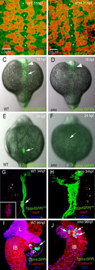
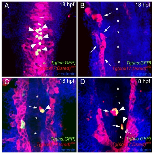
Absence of Dorsal Pancreatic Bud Derived β Cells in smo Mutants (A and B) Ventral confocal images of Tg(sox17:DsRed)s903 (red) and β-catenin (green) expression, comparing wild-type (A) and smo mutants (B) at 18 hpf (mid-trunk region). Tg(sox17:DsRed)s903-expressing endodermal cells show a similar arrangement and distribution around the midline (asterisks) in wild-type (A) and smo mutants (B). (C?F) Dorsal views of Tg(ins:GFP) embryos stained for GFP (green), comparing wild-type (C and E) and smo mutants (D and F) at 18 (C and D) and 24 (E and F) hpf. (C and D) Tg(ins:GFP)-expressing cells are completely missing in 18 hpf smo mutants. Cells in the anterior neural tube (arrowhead) also express the ins:GFP transgene. (E) Tg(ins:GFP)-expressing cells form a cluster in 24 hpf wild-type embryos (arrow). (F) Tg(ins:GFP)-expressing cells are completely missing in most smo mutants, though in approximately 5% of the mutants (n > 100 mutants examined), one or two Tg(ins:GFP)-expressing cells can be observed ([F], arrow). (G?J) Confocal projections of wild-type (G and I) and smo mutant (H and J) endoderm at 34 (G and H) and 96 (I and J) hpf. (G and H) Tg(gutGFP) s854 embryos were stained for Islet1 (red) and Insulin (blue). (G) The endodermal cells (green) form a solid multicellular rod, and pancreatic endocrine cells (arrow and inset) aggregate into one cluster in wild-type embryos. (H) In smo mutants, the endodermal cells fail to condense into a midline rod, and pancreatic endocrine cells are completely absent. (I and J) Tg(ins:GFP) embryos were stained for pan-Cadherin (red) and 2F11 (blue) to mark the endoderm and ductal structures, respectively. (I) Tg(ins:GFP)-expressing β cells (arrow) form a cluster inside the wild-type pancreas. (J) Although the morphology of the pancreas and liver is disrupted, Tg(ins:GFP)-expressing β cells (arrows) appear in smo mutants in ventral pancreatic bud derived tissues. L, liver; P, pancreas; IB, intestinal bulb. EXPRESSION / LABELING:
PHENOTYPE:
|

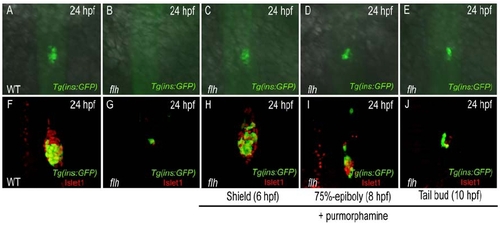
Shh Compensates for the Loss of Axial Mesoderm in the Formation of Pancreatic Endocrine Cells (A?C) Ventral confocal images of Tg(ins:GFP) (green) and Tg(sox17:DsRed)s903 (red) expression, comparing wild-type (A), flh mutants (B), and shh-overexpressing flh mutants (C) at 18 hpf. (C) At 18 hpf, Tg(ins:GFP)-expressing cells were rescued in shh-overexpressing flh mutants and scattered within the endodermal sheet (red). Most endodermal cells failed to converge following shh overexpression, leading to the presence of gaps (asterisks) within the endodermal sheet. (D?I) Lateral (D?F) and dorsal (G?I) views of Tg(ins:GFP) embryos comparing wild-type (D and G), flh mutants (E and H), and shh-overexpressing flh mutants (F and I) at 24 hpf. (D and G) Wild-type embryos show one cluster of Tg(ins:GFP)-expressing cells (arrow). (E and H) Tg(ins:GFP) expression is completely missing in flh mutants. (F and I) shh-overexpressing flh mutants show rescue of Tg(ins:GFP) expression (arrow). (J?O) Confocal projections of pancreatic endocrine cells comparing wild-type (J and M), flh mutants (K and N), and shh-overexpressing flh mutants (L and O) at 48 (J?L) and 60 (M?O) hpf. Tg(ins:GFP) embryos were stained for GFP (green), Somatostatin (blue), and Glucagon (red). flh mutants show dramatically reduced pancreatic endocrine cells at 48 (K) and 60 (N) hpf, compared to wild-type (J and M). Tg(ins:GFP) and Somatostatin expression were rescued at 48 hpf (L), and Glucagon expression was also detectable at 60 hpf (O) in shh-overexpressing flh mutants. EXPRESSION / LABELING:
|

Cell-Nonautonomous Requirement of Smoothened Function in the Induction of Dorsal Pancreatic Endocrine Cells (A) Schematic diagram of the cell transplantation protocol. cas-overexpressing wild-type or smo mutant Tg(ins:GFP) donor cells were transplanted into wild-type hosts. (B?G) Wild-type hosts with wild-type (B, D, and F) and smo mutant (C, E, and G) Tg(ins:GFP) donor cells at 30 hpf: dorsal views (B and C) and confocal projections (D?G). All donor cells are labeled with rhodamine dextran (red). Note that only donor cells can express the ins:GFP transgene. (B) Wild-type donor cells contribute to the endoderm and express the ins:GFP transgene in wild-type hosts (arrow; 52%, n = 77/147). (C) smo mutant donor cells contribute to the endoderm and express the ins:GFP transgene (arrow) in wild-type hosts as frequently as wild-type cells do (51%, n = 25/49). (D?G) Hosts were stained for Islet1, which marks all pancreatic endocrine cells (blue) and motoneurons (blue, asterisk). (D and F) Wild-type donor cells differentiate into Islet1-positive endocrine cells (red cytoplasm, blue nucleus; arrowhead), and many of the donor cells coexpress the ins:GFP transgene (triple-positive cells appear white; arrows). (E and G) smo mutant donor cells also differentiate into Islet1-positive endocrine cells (red cytoplasm, blue nucleus; arrowhead), and many of the donor cells coexpress the ins:GFP transgene (triple-positive cells appear white; arrows). Single- and double-channel images are shown in Figure S3. |
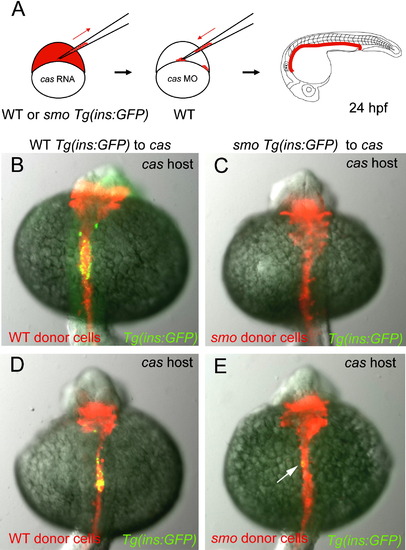
Smoothened Function Is Required in the Endoderm for the Induction of Dorsal Pancreatic β Cells (A) Schematic diagram of the cell transplantation protocol. cas-overexpressing wild-type or smo mutant Tg(ins:GFP) donor cells were transplanted into cas MO-injected wild-type hosts. (B?E) Dorsal views of the cas MO-injected hosts with wild-type (B and D) and smo mutant (C and E) donor cells at 24 hpf. Donor cells are labeled with rhodamine dextran (red). All the endodermal cells of the hosts are replaced by donor cells. (B and D) Wild-type-like Tg(ins:GFP) expression in the endoderm derived from wild-type donor cells (95%, n = 74/78). (C and E) Tg(ins:GFP)-expressing cells are completely absent ([C], 92%, n = 23/25) or amount to one or two cells ([E], arrow; 8%, n = 2/25) when smo mutant donor cells form the endoderm in cas MO-injected wild-type hosts. |

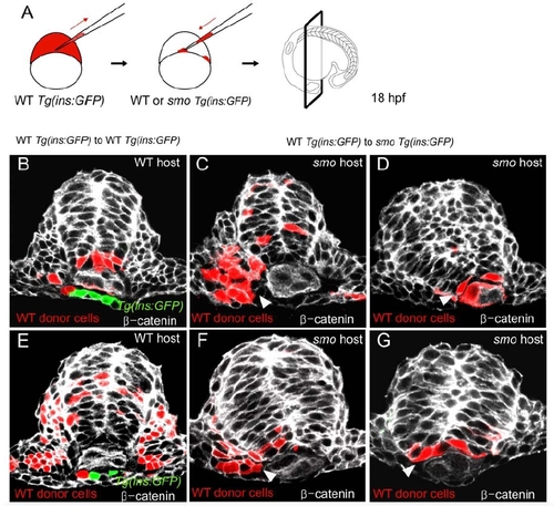
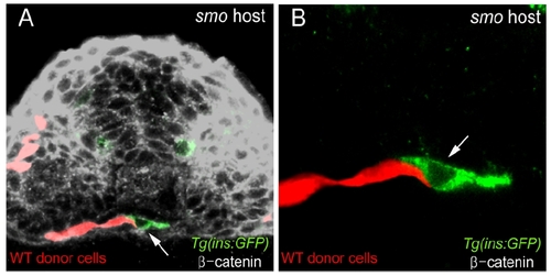
Cell-Cell Interactions between Endodermal Cells Are Required for Dorsal Pancreatic β Cell Induction (A) Schematic diagram of the cell transplantation protocol. cas-overexpressing wild-type Tg(ins:GFP) donor cells were transplanted into wild-type or smo mutant Tg(ins:GFP) hosts. (B and C) Dorsal views of wild-type (B) and smo mutant (C) hosts with the endodermal cells derived from wild-type donor cells labeled with rhodamine dextran (red) at 18 hpf. (B) Wild-type hosts show wild-type-like Tg(ins:GFP) expression after transplantation. (C) Tg(ins:GFP) expression (arrows) is restored in smo mutant hosts when wild-type cells contribute to the endoderm (44.5%, n = 48/108). (D and E) Transverse confocal images of smo mutant hosts stained for GFP (green) and β-catenin (white) at 18 hpf. Wild-type donor cells are labeled with rhodamine dextran (red). At this stage, parts of the endodermal tissue have become multilayered as cells are converging actively to the midline. (D) Most of the Tg(ins:GFP)-expressing cells in smo mutant hosts are derived from wild-type donor cells (arrows). (E) However, in several embryos (n = 6/48), lateral endoderm derived from wild-type donor cells correlates with Tg(ins:GFP) expression in medially located smo mutant host endoderm (arrow). (F) Schematic diagram of the cell transplantation protocol. cas-overexpressing wild-type donor cells were transplanted into wild-type or smo mutant Tg(ins:GFP) hosts. (G and H) Lateral views of live smo mutant hosts for Tg(ins:GFP) (green) and rhodamine dextran (red) at 30 hpf. (G) When wild-type donor cells were not incorporated into the endoderm, smo mutants showed a complete absence of Tg(ins:GFP) expression in most cases (48/50) or at most 2 Tg(ins:GFP)-expressing cells (2/50). (H) When wild-type donor cells were incorporated into the endoderm, several experimental smo mutants (n = 7/26) showed 4?7 rescued Tg(ins:GFP)-expressing cells (arrows; 5 rescued cells in this figure). (In these experimental smo mutants, pancreatic β cells likely comprise both wild-type and smo mutant cells.) |
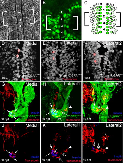
Lineage-Tracing Experiments Reveal that Endodermal Precursors Contribute to Distinct Endodermal Tissues Based on Their Medio-Lateral Position (A and B) Dorsal views of live Tg(sox17:GFP)s870 embryos at the 6- to 8-somite stage. (A) Bright-field image showing the region between somites 1 and 4. (B) Tg(sox17:GFP) expression shows that endodermal cells are spread in a monolayer and have not yet fused at the midline. (C) Schematic diagram of Tg(sox17:GFP)-expressing cells with three specific positions in the medio-lateral axis: the most medial cells (medial, dark green), cells immediately adjacent to the medial cells (lateral 1, green), and cells one cell apart from the medial cells (lateral 2, light green). Brackets indicate the area where uncaging was performed. (D?F) Ventral confocal images of control embryos at the 10-somite stage, confirming the accuracy of endodermal targeting by the laser. Tg(sox17:GFP)s870 embryos were stained for GFP (gray) and uncaged fluorescein (red). Two endodermal cells in the medial (D), lateral 1 (E), and lateral 2 (F) positions were labeled in the region between somites 2 and 3. (G?L) Confocal projections of Tg(sox17:GFP)s870 embryos at 50 hpf showing the progeny of the medial (G and J), lateral 1 (H and K), and lateral 2 (I and L) cells. Tg(sox17:GFP)s870 embryos were stained for GFP (green; [G?I]), Insulin (blue; [G?L]), and uncaged fluorescein (red; [G?L]). (G) and (J), (H) and (K), and (I) and (L) are the same embryos. (G and J) Medial cells gave rise exclusively to pancreatic endocrine cells (arrows). (H and K) Lateral 1 cells gave rise to exocrine cells (double arrows), a small number of endocrine cells (arrows), as well as intestinal tissue (arrowhead) adjacent to the pancreas. (I and L) Lateral 2 cells gave rise to liver cells (asterisks), intestine (arrowhead), and exocrine cells (double arrow). Lateral 2 cells showed very little, if any, contribution to pancreatic endocrine cells (arrow). EXPRESSION / LABELING:
|
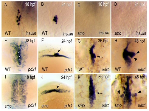
Absence of Dorsal Pancreatic Bud Derived β-Cells in smoothened (smo) Mutant Embryos EXPRESSION / LABELING:
|
Hedgehog Signaling Is Required during Gastrulation for Inducing Dorsal Pancreatic β-Cells EXPRESSION / LABELING:
|
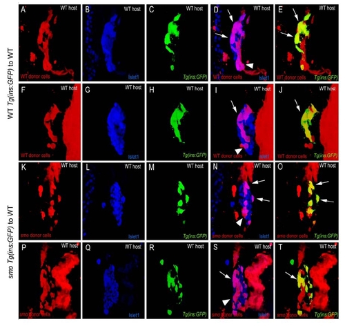
Cell Non-Autonomous Requirement of Smoothened Function in the Induction of Dorsal Pancreatic Endocrine Cells |
Smoothened Function in Adaxial and Lateral Floor Plate Cells Does Not Appear to Be Required for the Induction of Dorsal Pancreatic β-Cells |
Induction of Tg(ins:GFP) Expression in smo Mutants Is Coincidental with Close Contact with Wildtype Endodermal Cells |
Direct Endodermal Cell-Cell Contacts May Not Be Essential for Tg(ins:GFP) Expression EXPRESSION / LABELING:
|
Reprinted from Developmental Cell, 14(4), Chung, W.S., and Stainier, D.Y., Intra-endodermal interactions are required for pancreatic beta cell induction, 582-593, Copyright (2008) with permission from Elsevier. Full text @ Dev. Cell