
|
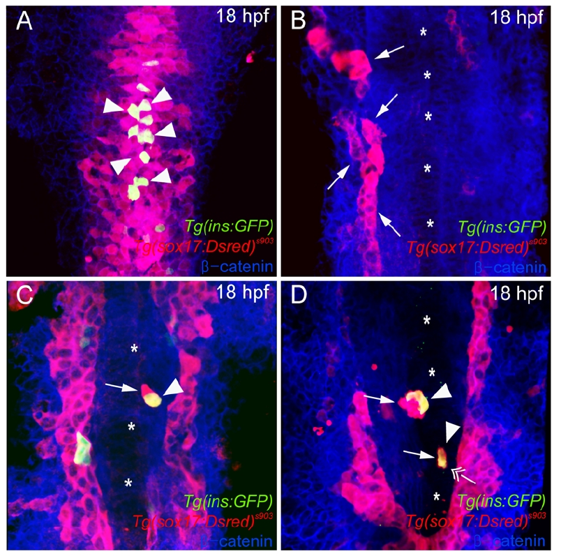
Fig. S6
Direct Endodermal Cell-Cell Contacts May Not Be Essential for Tg(ins:GFP) Expression
(A-B) Ventral confocal images of Tg(ins:GFP) (green), Tg(sox17:DsRed)s903 (red) and β- catenin (blue) expression, comparing wildtype (A) and cas MO (0.1∼0.4 ng) injected (B-D) embryos at 18 hpf (mid-trunk region). (A) In wildtype embryos, Tg(sox17:DsRed)s903 expressing endodermal cells are condensed near the midline and Tg(ins:GFP) expressing cell are located medially (arrowheads). (B) A majority (n = 53/67) of cas MO injected embryos did not exhibit Tg(ins:GFP) expression and showed a much reduced number of Tg(sox17:DsRed)s903 expressing cells. These endodermal cells (arrows) were located away from the midline (asterisks). (C-D) In the other 14 embryos, a few endodermal cells (arrows) had migrated close to the midline (asterisks) and were expressing the ins:GFP transgene (arrowheads). (D) Interestingly, in 4 such embryos, a single, isolated Tg(ins:GFP) expressing cell was observed (double arrow). Examining a set of 14-somite stage embryos led to similar observations (Table S4). It is interesting to note that in these experimental embryos, Tg(ins:GFP) expression was mostly observed close to the midline, possibly revealing the importance of medio-lateral information in the dorsal pancreatic β- cell induction process.
Reprinted from Developmental Cell, 14(4), Chung, W.S., and Stainier, D.Y., Intra-endodermal interactions are required for pancreatic beta cell induction, 582-593, Copyright (2008) with permission from Elsevier. Full text @ Dev. Cell