
Fig. 7
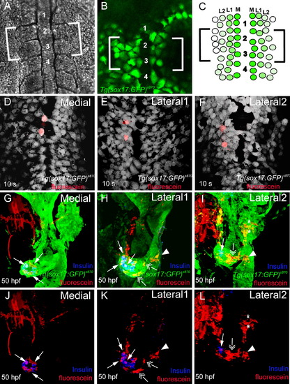
Lineage-Tracing Experiments Reveal that Endodermal Precursors Contribute to Distinct Endodermal Tissues Based on Their Medio-Lateral Position (A and B) Dorsal views of live Tg(sox17:GFP)s870 embryos at the 6- to 8-somite stage. (A) Bright-field image showing the region between somites 1 and 4. (B) Tg(sox17:GFP) expression shows that endodermal cells are spread in a monolayer and have not yet fused at the midline. (C) Schematic diagram of Tg(sox17:GFP)-expressing cells with three specific positions in the medio-lateral axis: the most medial cells (medial, dark green), cells immediately adjacent to the medial cells (lateral 1, green), and cells one cell apart from the medial cells (lateral 2, light green). Brackets indicate the area where uncaging was performed. (D–F) Ventral confocal images of control embryos at the 10-somite stage, confirming the accuracy of endodermal targeting by the laser. Tg(sox17:GFP)s870 embryos were stained for GFP (gray) and uncaged fluorescein (red). Two endodermal cells in the medial (D), lateral 1 (E), and lateral 2 (F) positions were labeled in the region between somites 2 and 3. (G–L) Confocal projections of Tg(sox17:GFP)s870 embryos at 50 hpf showing the progeny of the medial (G and J), lateral 1 (H and K), and lateral 2 (I and L) cells. Tg(sox17:GFP)s870 embryos were stained for GFP (green; [G–I]), Insulin (blue; [G–L]), and uncaged fluorescein (red; [G–L]). (G) and (J), (H) and (K), and (I) and (L) are the same embryos. (G and J) Medial cells gave rise exclusively to pancreatic endocrine cells (arrows). (H and K) Lateral 1 cells gave rise to exocrine cells (double arrows), a small number of endocrine cells (arrows), as well as intestinal tissue (arrowhead) adjacent to the pancreas. (I and L) Lateral 2 cells gave rise to liver cells (asterisks), intestine (arrowhead), and exocrine cells (double arrow). Lateral 2 cells showed very little, if any, contribution to pancreatic endocrine cells (arrow). |
| Gene: | |
|---|---|
| Fish: | |
| Anatomical Term: | |
| Stage: | 5-9 somites |
Reprinted from Developmental Cell, 14(4), Chung, W.S., and Stainier, D.Y., Intra-endodermal interactions are required for pancreatic beta cell induction, 582-593, Copyright (2008) with permission from Elsevier. Full text @ Dev. Cell