
- Title
-
nil per os encodes a conserved RNA recognition motif protein required for morphogenesis and cytodifferentiation of digestive organs in zebrafish
- Authors
- Mayer, A.N. and Fishman, M.C.
- Source
- Full text @ Development
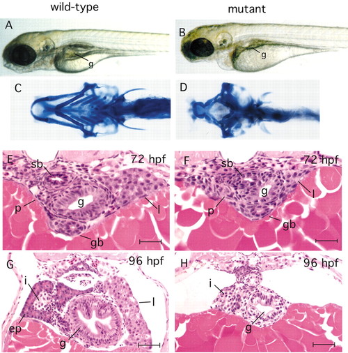
npo-mutant phenotype. (A,B) Live larvae at 4.5 dpf, showing that the jaw, branchial arches and gut tube are markedly underdeveloped at 96 hpf, but that dorsal structures (somites, notocord) appear unaffected by the mutation. (C,D) Alcian Blue staining of wild-type and mutant larvae at 4.5 dpf demonstrates failure of branchial arch growth beyond the initial primordium. (E-G) Cross sections of embryos stained with Hematoxylin and Eosin at the level of the pancreatic islet. (E,F) 72 hpf embryos (genotyped prior to embedding) showing the first morphologically detectable differences between mutant and wild type, with hypoplastic gut and liver in the mutant. (G,H) 96 hpf embryos show a stark contrast between wild type and mutant. The mutant gut tube is substantially smaller and the epithelial architecture is less organized, with no villi formation. The exocrine pancreas is not recognizable in the mutant, yet the islet appears relatively normal. The liver is not seen because it is much smaller in the AP dimension and does not extend to this level. g, gut; p, pancreas; l, liver; i, pancreatic islet; ep, exocrine pancreas; gb, gall bladder; sb, swim bladder. Scale bars: E,F, 20 μm; G,H, 30 μm. PHENOTYPE:
|
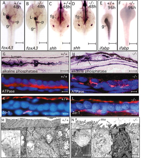
npo is required for endoderm-intestine transition. (A,B) foxa3 expression pattern at 48 hpf showing formation of liver, gut and pancreatic anlage in both wild-type and npo mutant embryos. (C,D) shh expression at 48 hpf reveals normal foregut patterning, with label exclusion at the prepancreatic endoderm. (E,F) ifabp expression detected in the wild-type intestine but not in the mutant. (G,H) Histochemical staining for alkaline phosphatase. Robust staining of apical aspect of epithelium in the wild type, but staining of mutant epithelial cells appears basal, suggesting mislocalization. (I,J), Na/K ATPase immunofluorescence demonstrates basolateral localization of fluorescence in wild type, but no clear localization in the mutant. (K,L) Zo1 immunofluorescence demonstrates formation of zona occludens in both wild-type and mutant embryos. (M,N) Transmission electron micrograph of intestine. Junctional complexes are present in both mutant and wild-type epithelia. Microvilli in the wild type are smooth and uniform, but in the mutant they are fewer and pleiomorphic. +/+, wild type; -/-, mutant; g, gut; L, liver; p, pancreas; mv, microvilli; TJ, tight junction. Scale bars: A-F, 150 μm; G,H, 20 μm; I-L, 10 μm; M,N, 1 μm; insets, 30 μm. |
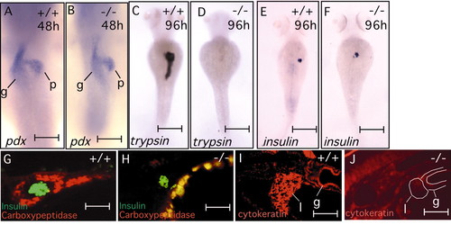
npo mediates exocrine pancreas and liver cytodifferentiation. (A-F) Whole-mount in situ hybridizations show formation of pancreatic primordia in both mutant and wild-type embryos at 48 hpf (A,B), and subsequent failure to form differentiated exocrine pancreas in mutant embryos by 96 hpf (C,D). By contrast, islet formation is independent of npo. (G,H), Double immunofluorescence staining for insulin (green) and carboxypeptidase (red) shows selective failure of exocrine pancreas formation in the mutant, with sparing of the pancreatic islet. (I,J) Immunofluorescence detection of cytokeratin (monoclonals AE1/AE3), demonstrating staining of wild-type liver and gut epilthelia, but absent specific staining of mutant epithelia. White outline defines organ boundaries identified by phase contrast. g, gut; p, pancreas; l, liver. Scale bars: A,B, 80 μm; C-E, 150 μm; G-J, 25 μm. |
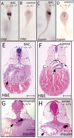
Rescue and phenocopy of npo mutation. (A,C,E) 96 hpf npo-mutant embryos that had been injected with NotI-digested BAC 37b12 at the 1-cell stage. (B,D,F) Mutants injected with BAC 37b12 pre-cut with restriction endonucleases NotI and SnaBI, which cuts only between the first and second exon of the npo gene. (G,H) Wild-type embryos injected with morpholino oligonucleotides encoding antisense and sense sequences, respectively, from the npo transcript. (A,B) Representative embryos stained by whole-mount in situ hybridization for ifabp show rescue of this marker by BAC 37b12 injection in the expected mosaic pattern, and undetectable rescue observed using the cleaved BAC. By comparing panel A with Fig. 2E, we note that the BAC-injected mutant embryos have a broader ifabp expression pattern than the wild-type embryos. (C,D) Staining for trypsin demonstrates mosaic rescue of the npo phenotype, also with a broader expression pattern than seen in the wild-type (compare with Fig. 3C). The control-injected embryos shows a few cells stained for trypsin expression, which we also note occasionally in control-uninjected embryos. (E,F) Embryos shown in A and B, respectively, were sectioned and stained with Hematoxylin and Eosin. This revealed an over-expanded intestinal and exocrine pancreatic epithelium in the embryos injected with intact BAC 37b12, but no histological effect on the mutant phenotype in the control (arrows). (G,H) Histological sections of embryo injected with npo-specific morpholino oligonucleotides, sense or antisense as indicated, stained for insulin by whole-mount in situ hybridization, then sectioned and counterstained with nuclear Fast Red. Antisense injection leads to hypoplasia, dysmorphogenesis and abrogation of epithelial cytodifferentiation similar to that seen in the npo-homozygous mutant. insulin expression is not affected, as seen in the homozygous mutant. Normal digestive organs are seen in the sense control. Note, the effects on the digestive organs due to manipulation of npo expression often result in uncoupling of the AP axes of the neural tube from the digestive organs. For this reason, we chose internal landmarks in the digestive tract (i.e. islet or gall bladder) rather than neural tube or somite landmarks. Arrows indicate intestine. Scale bars: A-D, 200 μm; E-H, 20 μm. EXPRESSION / LABELING:
|
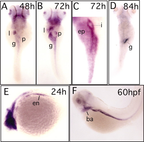
Developmental expression pattern of npo. (A-F) Whole-mount in situ hybridization to npo at the indicated developmental stages. (A,B,D) Dorsal view demonstrates dynamic expression in the digestive organ primordia. Expression begins at about 48 hpf, peaks at 72 hpf, then recedes in an anterior to posterior wave around 84 hpf. (C) Right oblique view showing expression of npo in the exocrine pancreas at 72 hpf, but not in the pancreatic islet. (E,F) Lateral view showing brain and endoderm expression at 24 hpf, then branchial arch and endoderm expression at 60 hpf. l, liver; g, gut; p, pancreas; i, islet; ep, exocrine pancreas; en, endoderm; ba, branchial arches. EXPRESSION / LABELING:
|
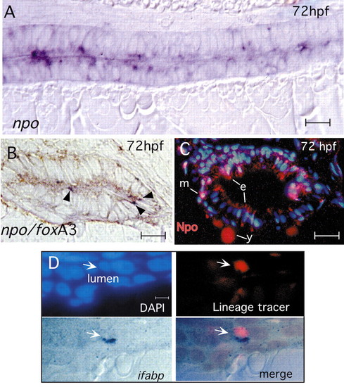
Expression and function of npo in the endoderm. (A) Mid-sagittal section of 72 hpf embryo labeled for npo by in situ hybridization, showing punctate, heterogeneous staining in epithelial cells. (B) Double in situ hybridization to npo (dark blue; arrowheads) and foxa3 (brown), showing npo expression in a subset of endoderm-derived epithelial cells. (C) Anti-Npo immunofluorescence (red) at 72 hpf shows expression primarily in gut epithelium, and also in scattered subepithelial mesenchymal cells. The yolk signal is caused by autofluorescence. (D) Mosaic analysis: wild-type cell (arrow) in npo-/- host shows cell autonomous rescue of ifabp expression. Panels show nuclei (DAPI; blue), lineage tracer (rhodamine dextran; red) and labeling for ifabp (dark blue). e, epithelium; m, mesenchyme; y, yolk. Scale bars: A,B, 10 μm; C, 15 μm; D, 7 μm. EXPRESSION / LABELING:
|

Unillustrated author statements |