
|
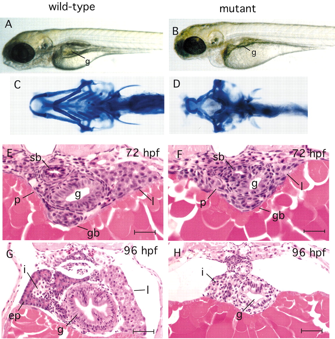
Fig. 1 npo-mutant phenotype. (A,B) Live larvae at 4.5 dpf, showing that the jaw, branchial arches and gut tube are markedly underdeveloped at 96 hpf, but that dorsal structures (somites, notocord) appear unaffected by the mutation. (C,D) Alcian Blue staining of wild-type and mutant larvae at 4.5 dpf demonstrates failure of branchial arch growth beyond the initial primordium. (E-G) Cross sections of embryos stained with Hematoxylin and Eosin at the level of the pancreatic islet. (E,F) 72 hpf embryos (genotyped prior to embedding) showing the first morphologically detectable differences between mutant and wild type, with hypoplastic gut and liver in the mutant. (G,H) 96 hpf embryos show a stark contrast between wild type and mutant. The mutant gut tube is substantially smaller and the epithelial architecture is less organized, with no villi formation. The exocrine pancreas is not recognizable in the mutant, yet the islet appears relatively normal. The liver is not seen because it is much smaller in the AP dimension and does not extend to this level. g, gut; p, pancreas; l, liver; i, pancreatic islet; ep, exocrine pancreas; gb, gall bladder; sb, swim bladder. Scale bars: E,F, 20 μm; G,H, 30 μm.