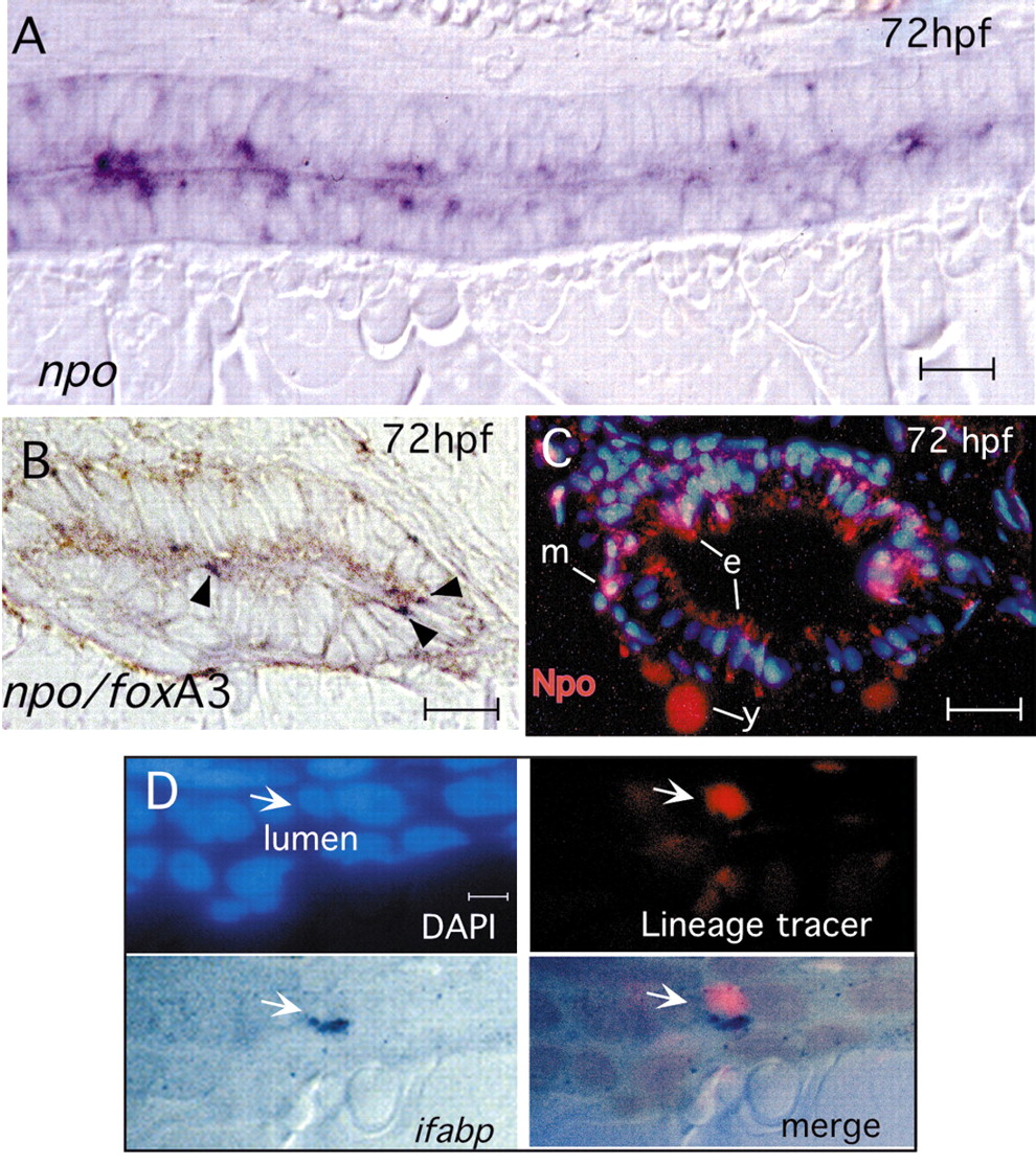
Fig. 8 Expression and function of npo in the endoderm. (A) Mid-sagittal section of 72 hpf embryo labeled for npo by in situ hybridization, showing punctate, heterogeneous staining in epithelial cells. (B) Double in situ hybridization to npo (dark blue; arrowheads) and foxa3 (brown), showing npo expression in a subset of endoderm-derived epithelial cells. (C) Anti-Npo immunofluorescence (red) at 72 hpf shows expression primarily in gut epithelium, and also in scattered subepithelial mesenchymal cells. The yolk signal is caused by autofluorescence. (D) Mosaic analysis: wild-type cell (arrow) in npo-/- host shows cell autonomous rescue of ifabp expression. Panels show nuclei (DAPI; blue), lineage tracer (rhodamine dextran; red) and labeling for ifabp (dark blue). e, epithelium; m, mesenchyme; y, yolk. Scale bars: A,B, 10 μm; C, 15 μm; D, 7 μm.
Image
Figure Caption
Figure Data
Acknowledgments
This image is the copyrighted work of the attributed author or publisher, and
ZFIN has permission only to display this image to its users.
Additional permissions should be obtained from the applicable author or publisher of the image.
Full text @ Development

