
|
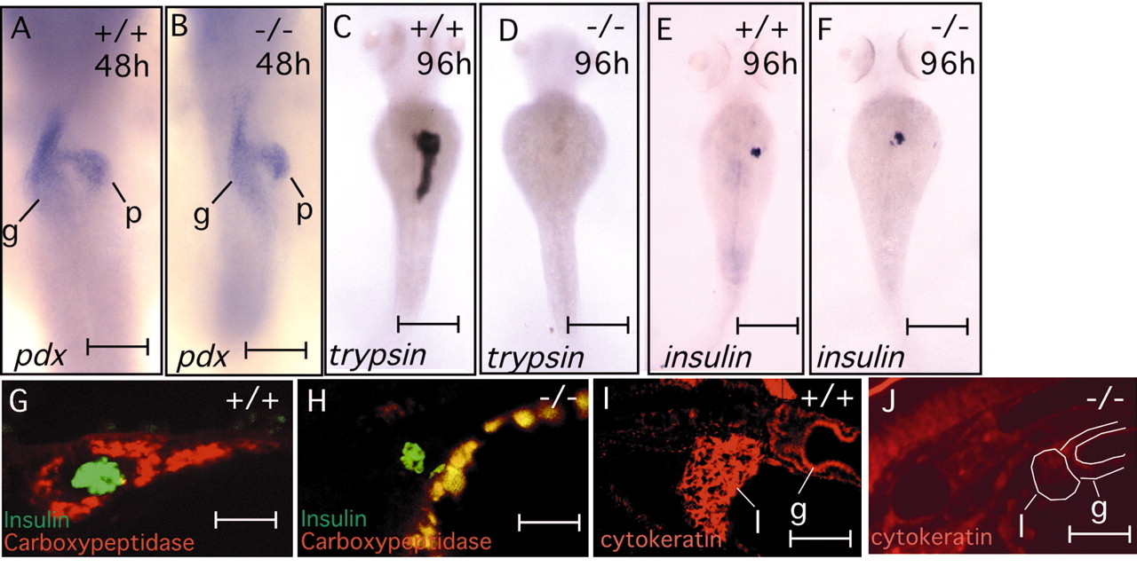
Fig. 3 npo mediates exocrine pancreas and liver cytodifferentiation. (A-F) Whole-mount in situ hybridizations show formation of pancreatic primordia in both mutant and wild-type embryos at 48 hpf (A,B), and subsequent failure to form differentiated exocrine pancreas in mutant embryos by 96 hpf (C,D). By contrast, islet formation is independent of npo. (G,H), Double immunofluorescence staining for insulin (green) and carboxypeptidase (red) shows selective failure of exocrine pancreas formation in the mutant, with sparing of the pancreatic islet. (I,J) Immunofluorescence detection of cytokeratin (monoclonals AE1/AE3), demonstrating staining of wild-type liver and gut epilthelia, but absent specific staining of mutant epithelia. White outline defines organ boundaries identified by phase contrast. g, gut; p, pancreas; l, liver. Scale bars: A,B, 80 μm; C-E, 150 μm; G-J, 25 μm.