
|
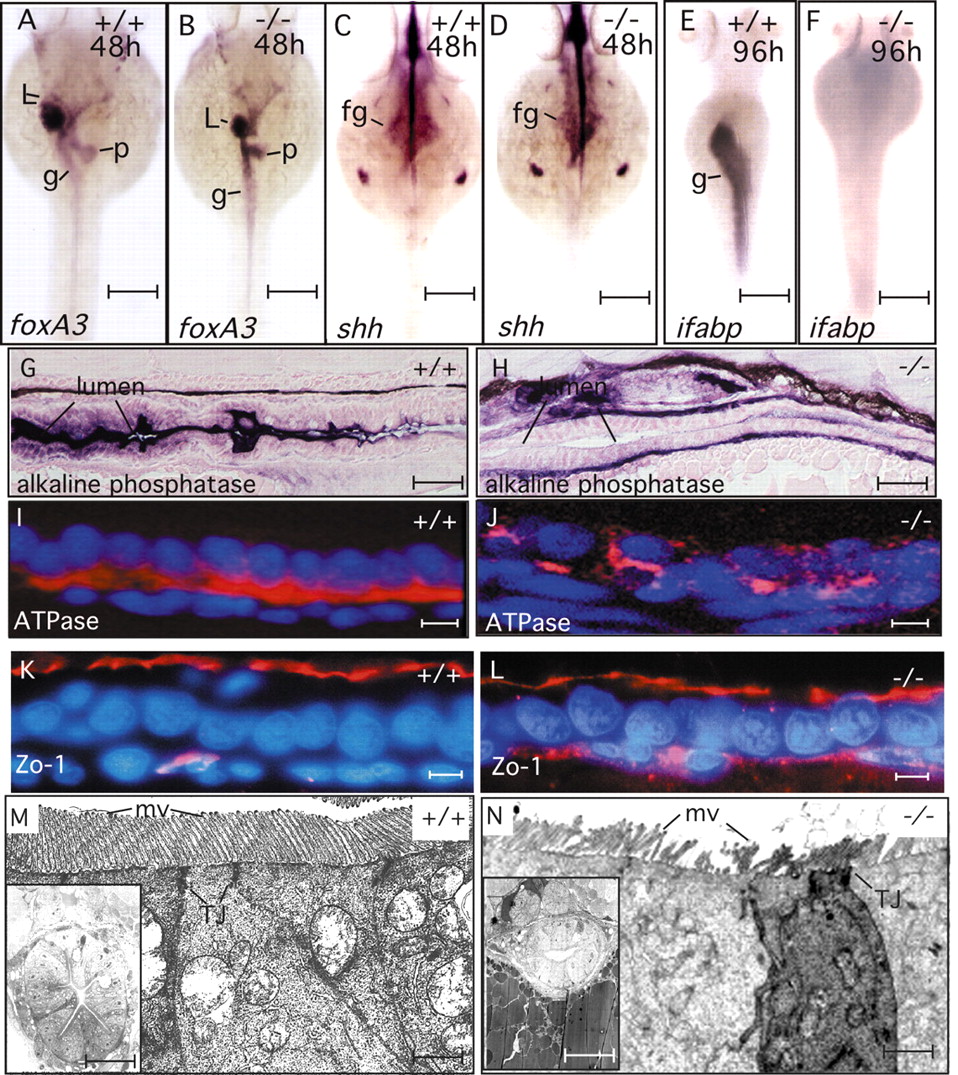
Fig. 2 npo is required for endoderm-intestine transition. (A,B) foxa3 expression pattern at 48 hpf showing formation of liver, gut and pancreatic anlage in both wild-type and npo mutant embryos. (C,D) shh expression at 48 hpf reveals normal foregut patterning, with label exclusion at the prepancreatic endoderm. (E,F) ifabp expression detected in the wild-type intestine but not in the mutant. (G,H) Histochemical staining for alkaline phosphatase. Robust staining of apical aspect of epithelium in the wild type, but staining of mutant epithelial cells appears basal, suggesting mislocalization. (I,J), Na/K ATPase immunofluorescence demonstrates basolateral localization of fluorescence in wild type, but no clear localization in the mutant. (K,L) Zo1 immunofluorescence demonstrates formation of zona occludens in both wild-type and mutant embryos. (M,N) Transmission electron micrograph of intestine. Junctional complexes are present in both mutant and wild-type epithelia. Microvilli in the wild type are smooth and uniform, but in the mutant they are fewer and pleiomorphic. +/+, wild type; -/-, mutant; g, gut; L, liver; p, pancreas; mv, microvilli; TJ, tight junction. Scale bars: A-F, 150 μm; G,H, 20 μm; I-L, 10 μm; M,N, 1 μm; insets, 30 μm.