
Fig. S19
- ID
- ZDB-FIG-170619-22
- Publication
- Minchin et al., 2017 - A classification system for zebrafish adipose tissues
- Other Figures
- All Figure Page
- Back to All Figure Page
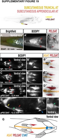
Morphology of abdominal SAT (ASAT) and SAT associated with the pelvic fin (PELSAT). A-A''. Brightfield (A), BODIPY (A') and false coloured BODIPY (A'') images to illustrate the earliest stages of PELSAT formation (arrows in A', and red in A''). B-D. BODIPY timeseries to illustrate the progressive growth of PELSAT (arrows) and ASAT. B'-D'. Magnified images to illustrate the wedge-shaped PELSAT and single streak of ASAT running along the ventral midline. B''-D''. False coloured BODIPY images based on B'-D' to show PELSAT (red) and ASAT (yellow). E. Lateral view to illustrate the morphology of PELSAT (red arrow) and ASAT (yellow arrow). The blue arrow indicates CVAT. F. At the location of the pelvic girdle, a discontinuous join is present in ASAT. G. Schematic illustrating the morphological characteristics of ASAT, PELSAT and CVAT. Scale bars are 1 mm (A-D), 500 μm (E). |