
FIGURE
Fig. S13
- ID
- ZDB-FIG-170619-16
- Publication
- Minchin et al., 2017 - A classification system for zebrafish adipose tissues
- Other Figures
- All Figure Page
- Back to All Figure Page
Fig. S13
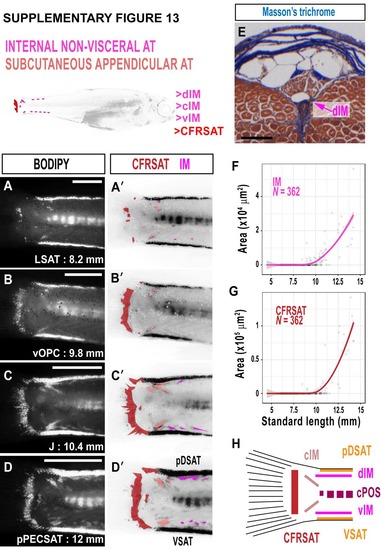
Intermuscular NVAT (IM) and caudal fin ray SAT (CFRSAT) in zebrafish. A-D. BODIPY timeseries illustrating the growth of IM and CFRSAT in the zebrafish tail. A'-D'. False coloured BODIPY images derived from A-D. CFRSAT is dark red, and IM deposits are magenta. E. Masson's trichrome-stained cross-section through the adult zebrafish tail showing dorsal IM (dIM). F. Relationship between IM and SL. G. Relationship between CFRSAT and SL. H. Schematic illustrating the multiple ATs within the zebrafish tail. Scale bars are 500 μm (A-D) and 100 μm (E). Fitted lines were generated by a LOESS function. |
Expression Data
Expression Detail
Antibody Labeling
Phenotype Data
Phenotype Detail
Acknowledgments
This image is the copyrighted work of the attributed author or publisher, and
ZFIN has permission only to display this image to its users.
Additional permissions should be obtained from the applicable author or publisher of the image.
Full text @ Dis. Model. Mech.