
- Title
-
Loss of mpv17 affected early embryonic development via mitochondria dysfunction in zebrafish
- Authors
- Bian, W.P., Pu, S.Y., Xie, S.L., Wang, C., Deng, S., Strauss, P.R., Pei, D.S.
- Source
- Full text @ Cell Death Discov
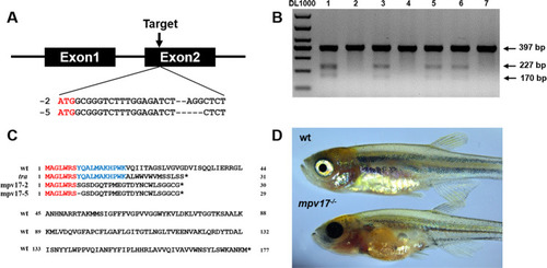
A The targeted loci were near the translation start code ATG of the gene mpv17. B The agarose gel electrophoresis results of the T7E1 assay for the screening of mpv17 knocked out zebrafish. The marker is DL1000 and the lane number shows different samples. The 397 bp band indicates the wild-type gene, and the lanes with 227 and 170 bp bands show the mutated genes. C The amino acid alignment of the mpv17 protein sequence in wild-type, tra mutant, and mpv17−/− zebrafish in this study. D One-month-old wild-type and mpv17−/− zebrafish larvae. PHENOTYPE:
|
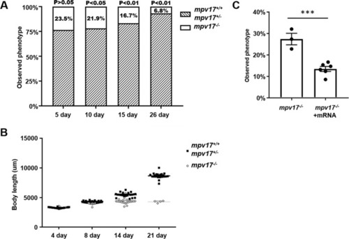
PHENOTYPE:
|
PHENOTYPE:
|
|
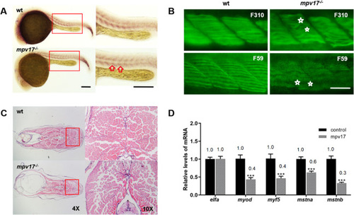
EXPRESSION / LABELING:
PHENOTYPE:
|
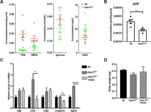
PHENOTYPE:
|