FIGURE SUMMARY
Title

Marcksb plays a key role in the secretory pathway of zebrafish Bmp2b

Authors
Ye, D., Wang, X., Wei, C., He, M., Wang, H., Wang, Y., Zhu, Z., Sun, Y.
Source
Full text @ PLoS Genet.

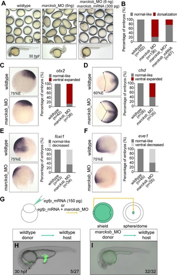
Marcksb is required for specification of ventral cell fate.

(A) Knockdown of marcksb showed dorsalization defects, which could be rescued by overexpression of morpholino insensitive mRNA of marcksb. Up panels, field view of embryos at early-somite stage; lower panels, representative embryos at 24 hours post fertilization (hpf). (B) The percentage of embryos with normal-like or dorsalization defects. “n” represents the number of embryos we observed. (C-F) Whole-mount in situ hybridization (WISH) showed the expansion of dorsal markers otx2 (neural ectoderm) (C) and chordin (dorsal margin) (D) expression, and the shrinkage of ventral markers foxi1 (non-neural ectoderm) (E) and eve1 (ventral margin) (F) expression in marcksb morphants. The percentage of embryos with indicated phenotype were shown aside their representative images. For otx2, foxi1 and eve1, embryos are lateral view with animal-pole to the top and dorsal to the right; for chd, embryos are animal-pole view with dorsal to the right; arrows in chd panels indicate the expansion locations of signals. (G) A schematic showing the procedure of the tail organizer transplantation assay. (H) A representative wildtype-to-wildtype transplantation embryo showing an induction of ectopic tail by grafting the tail organizer region of the wildtype embryo. (I) A representative morphant-to-wildtype transplantation embryo without induction of ectopic tissue by grafting the tail organizer region of the marcksb morphant embryo. The ratio at the right corner indicates the number of embryos with the representative phenotype/the total number of observed embryos.

Marcksb is required for activation of BMP signaling.

(A) WISH analysis showed that the transcriptional levels of downstream targets of BMP signaling—szl and ved were decreased in marcksb morphants. (B) The percentage of embryos with normal or decreased expression. “n” represents the number of embryos we observed. (C) The overall intensity of p-Smad1/5/9 at ventral region in marcksb morphants was dramatically decreased when compared with wildtype. (D) The normalized fluorescent intensity of P-Smad1/5/9. Error bars of light blue and light red show S.E.M. (E) Knockdown of marcksb could partially rescue the ventralization phenotype caused by injection of bmp2b mRNA (5 pg bmp2b mRNA per embryo). The statistical data are shown in the bar graphs with the number of observed embryos. “n” represents the number of embryos we observed. (F) Overexpression of bmp2b caused dramatic dorsal expansion of szl and ved expression while knockdown of marcksb in the bmp2b-overexpressed embryos partially restored their expression patterns. (G) The percentage of embryos with robust-increased or dorsal-inhibited expression. “n” represents the number of embryos we observed.

EXPRESSION / LABELING:
Genes:
Antibody:
Fish:
Knockdown Reagent:
Anatomical Term:
Stage: Shield
PHENOTYPE:
Fish:
Knockdown Reagent:
Observed In:
Stage: Shield

Phosphorylation and de-phosphorylation of Marcksb is required for the activation of BMP signaling.

(A) Overexpression of marcksb at high dosage (1000 pg per embryo) caused ventralization defects. “asterisk” shows the notochord; “arrow” indicates the disappearance of notochord; “arrow head” indicates the enlarged blood island. (B) The percentage of embryos with normal-like, moderate ventralization or severe ventralization. “n” represents the number of embryos we observed. (C) Phosphorylation and de-phosphorylation mutation types of Marcksb altered the sub-cellular location of Marcksb. (C-a) a diagram showing mutated regions of S4D-Marcksb and S4N-Marcksb; (C-b) A schematic showing the procedure of mosaic overexpression assay; (C- c, d and e) confocal microscopy analysis at shield stage showed that wildtype Marcksb and S4N-Marcksb co-localized with memGFP while S4D-Marcksb did not. (D) Representative images showing the expression of szl in wildtype embryos (D-a), and the embryos injected with marcksb mRNA (D-b), S4D-marcksb mRNA (D-c) and S4N-marcksb mRNA (D-d) at shield stage. Embryos are animal-pole view with dorsal to the right. (E) The percentage of embryos with normal-like, decreased or mildly increased expression of szl in different experimental groups indicated in the (D). “n” represents the number of embryos we observed. (F, G) Genetic interaction was examined by co-injection of sub-dose marcksb mRNA (200 pg/embryo) and chd_MO. (F) representative embryos showing for phenotypes of normal-like, moderate ventralization (V1-V2, ventralization type 1 to type 2) and severe ventralization (V3-V4, ventralization type 3 to type 4); (G) The percentage of embryos with normal-like, moderate ventralization or severe ventralization. “n” represents the number of embryos we observed. (H) Representative images showing the expression of szl in the shield-stage embryos injected with chd_MO (H-a), chd_MO plus marcksb mRNA (H-b), chd_MO plus S4D-marcksb mRNA (H-c), and chd_MO plus S4N-marcksb (H-d). Embryos are animal-pole view with dorsal to the right. (I) The percentage of embryos with rescued, increased or dramatically increased expression of szl in different experimental groups indicated in the (H). “n” represents the number of embryos we observed.

Marcksb cell autonomously regulates the extracellular level of Bmp2b.

(A) Injection of either myc-bmp2b or mcherry-bmp2b caused severe ventralization at 24 hpf, and injection of mcherry-bmp2b could rescue the dorsalization of bmp2b mutant. (A-a) wildtype embryos; (A-b) myc-bmp2b injected embryos; (A-c) mcherry-bmp2b injected embryos; (A-d) bmp2b mutant (allele name: bmp2bta72a/ta72a); (A-e) representative imaging showing mcherry-bmp2b rescued bmp2bta72a/ta72a; (A-f) representative imaging showing ventralized bmp2bta72a/ta72a by over-dosage of mcherry-bmp2b. All embryos were at 24 hpf. (B) The pCS2-myc-bmp2b plasmid was transfected into 293T cells. Intracellular and extracellular Myc-Bmp2b were analyzed by immunoblotting using anti-Myc antibody. The positions of precursor and mature Bmp2b are illustrated schematically to the right of each gel. (C) The pCS2-mcherry-bmp2b plasmid and pCS2-mcherry plasmid were transfected into 293T cells separately. Intracellular and extracellular mCherry-Bmp2b or mCherry were analyzed by immunoblotting using anti-mCherry antibody. The positions of mCherry-Bmp2b precursor, mature mCherry-Bmp2b and mCherry alone are illustrated schematically to the right of each gel. (D) Knockdown of marcksb reduced the extracellular level of Bmp2b. (D-a) showed a diagram of mosaic injection; (D-b) showed the extracellular level of mCherry-Bmp2b in wildtype embryos; (D-c) showed the extracellular level of mCherry-Bmp2b was significantly reduced in the marcksb morphant embryos. (E) Quantitative measurement of secreted Bmp2b in wildtype and marcksb morphant embryos. The data were presented as scatter plots with median; *”: P < 0.01, from Student’s t-test. (F) The mCherry-bmp2b mRNA injected wildtype embryos and marcksb morphants were collected separately at shield stage. Total cell-derived and extracellular mCherry-Bmp2b were analyzed by immunoblotting using anti-mCherry antibody. The positions of mCherry-Bmp2b precursor and mature mCherry-Bmp2b are illustrated schematically to the right of each gel. (G) Assay on secreted Myc-Bmp2b of wildtype cells and marcksb-depleted cells by transplantation. (G-a) A diagram of cell transplantation; (G-b, c, d) The transplanted embryos at shield stage, indicating the location of transplanted cell populations: ventral (b), lateral (c) and dorsal (d); (G-e, f, g) When wildtype donor cells were transplanted into wildtype host, the extracellular Myc-Bmp2b were at a comparable level among ventral (e), lateral (f) and dorsal transplants (g); (G-h) When wildtype donor cells were transplanted into the marcksb morphants, the extracellular Myc-Bmp2b was not reduced; (G-i) When the marcksb morphants cells were transplanted into wildtype host, the extracellular Myc-Bmp2b was strongly inhibited.

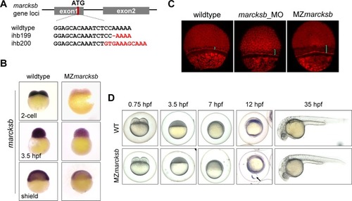
MZ<italic>marcksb</italic> do not show visible dorsoventral defects.

(A) The diagram shows the gRNA target of marcksb genome locus and the genotypes of two individual marcksb mutants generated by CRISPR/Cas9. (B) The transcription level of marcksb was significantly decreased in MZmarcksb embryos. Embryos of 2-cell stage, high stage and shield stage were present. All the embryos were lateral view with animal-pole to the top. (C) Both MZmarcksb and marcksb morphants showed similar defect of epiboly movement. The blue bar indicates the distance between the inner layer of cells and the envelope layer. (D) The wildtype and MZmarcksb embryos at developmental stages of 0.75 hpf, 3.5 hpf, 7 hpf, 12 hpf and 35hpf. MZmarcksb showed yolk bulge phenotype during early somite stage but normal appearance of development at 35 hpf. “Arrow” indicates the extrusion of the vegetal-most part of the yolk.

MZ<italic>marcksb</italic> showed genetic compensation on BMP signaling.

(A, B) Injection of marcksb_MO in MZmarcksb did not cause defect of dorsalization. (A) morphological observation of MZmarcksb and marcksb_MO injected MZmarcksb. Up panels, field view of embryos at 12 hpf; lower panels, representative embryos at 30 hpf. (B) WISH analysis showed that the transcriptional levels of downstream targets of BMP signaling—szl and ved were at comparable level between MZmarcksb and marcksb_MO injected MZmarcksb. (C) The overall intensity of p-Smad1/5/9 at ventral region in MZmarcksb was slightly increased when compared with wildtype. (D) The normalized fluorescent intensity of P-Smad1/5/9. Error bars of light blue and light red show S.E.M. (E) The extracellular mCherry-Bmp2b was slightly increased in the MZmarcksb compared with wildtype embryos. (F) Quantitative measurement of secreted Bmp2b in wildtype and MZmarcksb embryos. The data were presented as scatter plots with median; *”: P < 0.01, from Student’s t-test. (G) Although knockdown of chd in wildtype embryos only resulted in moderate ventralization (V1-V2), knockdown of chd in MZmarcksb embryos resulted in severe ventralization (V3-V4). V1-V4, ventralization type 1 to type 4 embryos; the statistical data are shown in the bar graphs with the number of observed embryos indicated right. (H) After injection of chd_MO, the up-regulation of BMP signaling activity was much more robust in MZmarcksb than that in wildtype embryos shown by WISH analysis of szl (a vs b) and ved (c vs d). The embryos are at shield stage and animal-pole view with dorsal to the right; (I) The percentage of embryos with normal-like, increased, and dramatically-increased expression of szl and ved. “n” represents the number of embryos we observed.

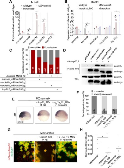
Up-regulation of the MARCKS family members and its interaction protein Hsp70.3 over-compensates the genetic loss of <italic>marcksb</italic>.

(A, B) RT-qPCR analysis on the expression of marcksa, marcksb, marcksl1a, marcksl1b and hsp70.3 in the embryos indicated in the Fig at 1-cell stage (A) and shield stage (B). The data were presented as scatter plots with bar representing median value relative to respective transcript levels measured in wildtype embryos. “*”: P < 0.01, from Student’s t-test. (C) Overexpression of marcksa, marcksl1a, marcksl1b or hsp70.3 partially rescued the dorsalization defects in marcksb morphants. “n” represents the number of embryos we observed. (D) Co-immunoprecipitation revealed that Hsp70.3 had high binding affinity with Marcksb and moderate binding affinity with Marcksl11a and Marcksl1b, but relative low binding affinity with Marcksa. TCL: total cell lysis. The molecular mass of Marcks-myc is around 70KD. (E) The expression of szl was remarkably reduced in MZmarcksb injected with either hsp70_MO or marcksa_l1a_l1b_MO. The embryos are at shield stage and lateral view with dorsal to the right. (F) The percentage of embryos with normal-like and severely decreased expression of szl. “n” represents the number of embryos we observed. (G) Hsp70 interacts with Marcksa, Marcksl1a and Marcksl1b to maintain sufficient level of extracellular Bmp2b in MZmarcksb. (a-c) Compared with MZmarcksb embryos (a), knockdown of either hsp70 (b) or a combination of marcksa, marcksl1a and marcksl1b (c) remarkably reduced the level of extracellular Bmp2b in MZmarcksb. (H) Quantitative measurement of secreted Bmp2b in embryos of MZmarcksb, MZmarcksb injected with either hsp70_MO or marcksa_l1a_l1b_MO. The data were presented as scatter plots with median; “*”: P < 0.01, from Student’s t-test.

Marcksb interacts with Hsp70.3 to regulate the dorsoventral patterning and the extracellular level of Bmp2b.

(A) Morphological phenotypes of embryos at 12 hpf, injected with sub-dosages of morpholinos against marcksb or hsp70 or the combination of both (marcksb_MO: 3 ng/embryo; hsp70_MO: 2 ng/embryo). (B-E) WISH analysis of szl (B), ved (C), otx2 (D), foxi1 (E). The percentage of embryos with different phenotypes for each group were indicated in the graph. Dosages of morpholino per embryo are indicated in the figure panel. For szl, ved and otx2, embryos are animal-pole view with dorsal to the right; for foxi1, embryos are lateral view with dorsal to the right. “n” represents the number of embryos we observed. (F) Quantitative measurement of secreted Bmp2b. The data were presented as scatter plots with median; “*”: P < 0.01, from Student’s t-test.

ZFIN is incorporating published figure images and captions as part of an ongoing project. Figures from some publications have not yet been curated, or are not available for display because of copyright restrictions.

EXPRESSION / LABELING:
Genes:
Fish:
Anatomical Term:
Stage Range: 2-cell to 75%-epiboly

ZFIN is incorporating published figure images and captions as part of an ongoing project. Figures from some publications have not yet been curated, or are not available for display because of copyright restrictions.

Acknowledgments
This image is the copyrighted work of the attributed author or publisher, and ZFIN has permission only to display this image to its users. Additional permissions should be obtained from the applicable author or publisher of the image. Full text @ PLoS Genet.