IMAGE

Fig 4

ID
ZDB-IMAGE-191230-1467
Source
Figures for Ye et al., 2019
Image
Figure Caption

Fig 4 Marcksb cell autonomously regulates the extracellular level of Bmp2b.

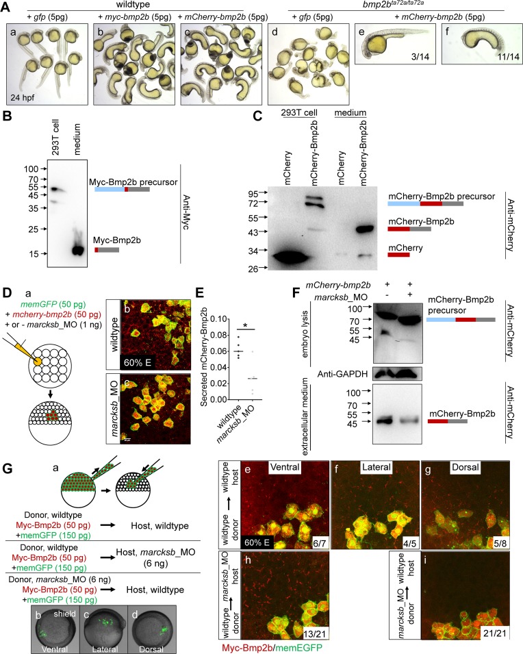
(A) Injection of either myc-bmp2b or mcherry-bmp2b caused severe ventralization at 24 hpf, and injection of mcherry-bmp2b could rescue the dorsalization of bmp2b mutant. (A-a) wildtype embryos; (A-b) myc-bmp2b injected embryos; (A-c) mcherry-bmp2b injected embryos; (A-d) bmp2b mutant (allele name: bmp2bta72a/ta72a); (A-e) representative imaging showing mcherry-bmp2b rescued bmp2bta72a/ta72a; (A-f) representative imaging showing ventralized bmp2bta72a/ta72a by over-dosage of mcherry-bmp2b. All embryos were at 24 hpf. (B) The pCS2-myc-bmp2b plasmid was transfected into 293T cells. Intracellular and extracellular Myc-Bmp2b were analyzed by immunoblotting using anti-Myc antibody. The positions of precursor and mature Bmp2b are illustrated schematically to the right of each gel. (C) The pCS2-mcherry-bmp2b plasmid and pCS2-mcherry plasmid were transfected into 293T cells separately. Intracellular and extracellular mCherry-Bmp2b or mCherry were analyzed by immunoblotting using anti-mCherry antibody. The positions of mCherry-Bmp2b precursor, mature mCherry-Bmp2b and mCherry alone are illustrated schematically to the right of each gel. (D) Knockdown of marcksb reduced the extracellular level of Bmp2b. (D-a) showed a diagram of mosaic injection; (D-b) showed the extracellular level of mCherry-Bmp2b in wildtype embryos; (D-c) showed the extracellular level of mCherry-Bmp2b was significantly reduced in the marcksb morphant embryos. (E) Quantitative measurement of secreted Bmp2b in wildtype and marcksb morphant embryos. The data were presented as scatter plots with median; *”: P < 0.01, from Student’s t-test. (F) The mCherry-bmp2b mRNA injected wildtype embryos and marcksb morphants were collected separately at shield stage. Total cell-derived and extracellular mCherry-Bmp2b were analyzed by immunoblotting using anti-mCherry antibody. The positions of mCherry-Bmp2b precursor and mature mCherry-Bmp2b are illustrated schematically to the right of each gel. (G) Assay on secreted Myc-Bmp2b of wildtype cells and marcksb-depleted cells by transplantation. (G-a) A diagram of cell transplantation; (G-b, c, d) The transplanted embryos at shield stage, indicating the location of transplanted cell populations: ventral (b), lateral (c) and dorsal (d); (G-e, f, g) When wildtype donor cells were transplanted into wildtype host, the extracellular Myc-Bmp2b were at a comparable level among ventral (e), lateral (f) and dorsal transplants (g); (G-h) When wildtype donor cells were transplanted into the marcksb morphants, the extracellular Myc-Bmp2b was not reduced; (G-i) When the marcksb morphants cells were transplanted into wildtype host, the extracellular Myc-Bmp2b was strongly inhibited.

Acknowledgments
This image is the copyrighted work of the attributed author or publisher, and ZFIN has permission only to display this image to its users. Additional permissions should be obtained from the applicable author or publisher of the image. Full text @ PLoS Genet.