|
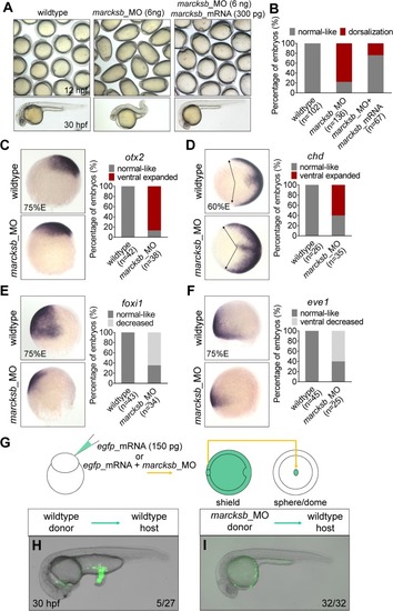
Marcksb is required for specification of ventral cell fate.(A) Knockdown of marcksb showed dorsalization defects, which could be rescued by overexpression of morpholino insensitive mRNA of marcksb. Up panels, field view of embryos at early-somite stage; lower panels, representative embryos at 24 hours post fertilization (hpf). (B) The percentage of embryos with normal-like or dorsalization defects. “n” represents the number of embryos we observed. (C-F) Whole-mount in situ hybridization (WISH) showed the expansion of dorsal markers otx2 (neural ectoderm) (C) and chordin (dorsal margin) (D) expression, and the shrinkage of ventral markers foxi1 (non-neural ectoderm) (E) and eve1 (ventral margin) (F) expression in marcksb morphants. The percentage of embryos with indicated phenotype were shown aside their representative images. For otx2, foxi1 and eve1, embryos are lateral view with animal-pole to the top and dorsal to the right; for chd, embryos are animal-pole view with dorsal to the right; arrows in chd panels indicate the expansion locations of signals. (G) A schematic showing the procedure of the tail organizer transplantation assay. (H) A representative wildtype-to-wildtype transplantation embryo showing an induction of ectopic tail by grafting the tail organizer region of the wildtype embryo. (I) A representative morphant-to-wildtype transplantation embryo without induction of ectopic tissue by grafting the tail organizer region of the marcksb morphant embryo. The ratio at the right corner indicates the number of embryos with the representative phenotype/the total number of observed embryos.
|