- Title
-
foxc1 is required for embryonic head vascular smooth muscle differentiation in zebrafish
- Authors
- Whitesell, T.R., Chrystal, P.W., Ryu, J.R., Munsie, N., Grosse, A., French, C.R., Workentine, M.L., Li, R., Zhu, L.J., Waskiewicz, A., Lehmann, O.J., Lawson, N.D., Childs, S.J.
- Source
- Full text @ Dev. Biol.
|
foxc1b:EGFP expressing mesenchymal cells attach to vessels and take on a vSMC morphology. A) Images of the ventral head in 2 dpf zebrafish embryos show foxc1b:EGFP positive mesenchymal cells near the endothelium (kdrl:mCherry; n = 19 embryos). B) At 3 dpf, foxc1b:EGFP positive cells associate with the endothelium (n = 14 embryos). Arrowheads indicate mural cells associated with the endothelium. C) At 4 dpf, foxc1b:EGFP positive cells flatten and tightly associate with the endothelium (n = 11 embryos). Scale bars represent 50 μm. |
|
Timelapse of foxc1b expressing mesenchymal cells attaching to the endothelium followed by upregulation of acta2. A) Ventral views of aortic arch arteries of a foxc1b:EGFP; kdrl:mCherry embryo using confocal time-lapse imaging starting at 50 hpf. Mesenchymal cells are indicated by the asterisk, while vascular mural cells are indicated by arrowheads, starting by 56–58 hpf. B) Orthogonal views at 58 hpf depict the foxc1b:EGFP cells wrapping around, but not co-expressing an endothelial marker. C) Time-lapse of the early ventral aorta of a foxc1b:EGFP; acta2:mCherry embryo from 60 to 70 hpf. foxc1b:EGFP positive smooth muscle cells are associated with the endothelium (arrowheads) but do not co-express acta2:mCherry until approximately 66 hpf (arrow). D) Single channel images of acta2:mCherry. E) Orthogonal views at 70 hpf show acta2:mCherry co-expressed within a foxc1b:EGFP positive cell. Scale bars represent 20 μm. |
|
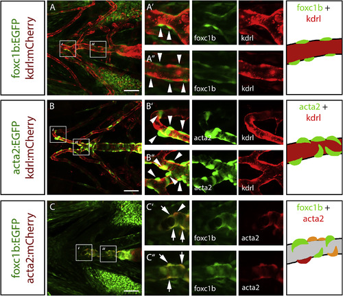
foxc1b expressing perivascular cells in the embryonic ventral head co-express acta2. A) In ventral head vessels at 4 dpf, foxc1b:EGFP expressing cells are associated with, and surround, the endothelium (kdrl:mCherry) along the ventral aorta. B) Similarly, acta2:EGFP positive smooth muscle cells surround the kdrl:mCherrypositive endothelium along the ventral aorta and aortic arch arteries. C) foxc1b:EGFP and acta2:mCherry are co-expressed along the ventral aorta. Schematics depict mural cell and endothelial marker expression matching the transgenes. Grey indicates presumptive endothelial patterns. Arrowheads represent marker-expressing mural cells. Arrows show co-expression of two smooth muscle cell markers. Scale bars represent 50 μm. EXPRESSION / LABELING:
|
|
foxc1b and acta2 are co-expressed in vSMCs in the adult zebrafish brain. A) foxc1b:EGFP expressing cells are closely associated with endothelial cells (kdrl:mCherry) in the adult brain. Arrowheads point to mural cells associated with the endothelium. Arrows point to co-expression of two smooth muscle cell markers. B) acta2:EGFPexpressing smooth muscle cells are closely associated with kdrl:mCherry expressing endothelial cells along large vessels of the brain. C) foxc1b:EGFP and acta2:mCherry are partially co-expressed in smooth muscle cells on brain vessels. Scale bars represent 50 μm. EXPRESSION / LABELING:
|
|
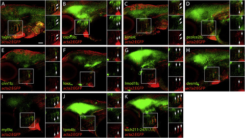
Expression of genes with enriched expression in the embryonic vSMC transcriptome. Gene expression from in situhybridization in laterally mounted 4 dpf embryos. Gene of interest mRNA is depicted in green, with αGFP antibody (red) detecting acta2:EGFP transgenic expression in smooth muscle cells. Arrows depict co-expression of gene of interest mRNA with acta2:EGFPexpression. A) tagln is expressed in the aortic arch arteries, and co-expressed with acta2:EGFP. B) capn3b expressed in the aortic arch arteries and brain. C) kcne4 is lowly expressed in the aortic arch arteries. D) pcolce2b is expressed in the aortic arch arteries and the brain. E) cnn1b is expressed in the aortic arch arteries. F) loxa is expressed in the aortic arch arteries, the bulbus arteriosus, and the ventral brain. G) lmod1b is expressed in the aortic arch arteries and brain. H) desmb is expressed in the aortic arch arteries, and in punctate patterns in the brain. I) myl9a is expressed in the aortic arch arteries. J) tpm4b is expressed in the brain and lowly expressed in the aortic arch arteries. K) si:ch211-247i17.1 is expressed in the aortic arch arteries and brain. Scale bar represents 50 μm. EXPRESSION / LABELING:
|
|
Necessity of foxc1 for embryonic head vSMC development. A) Ventral images of 3 dpf embryos depicting ventral aorta (red) and smooth muscle coverage (green) in foxc1a/foxc1b mutant genotypes. B) Ventral images of 4 dpf embryos depicting ventral aorta (red) and smooth muscle coverage (green) in foxc1a/foxc1b mutant genotypes. Genotypes are listed in each corresponding panel. C) At 4 dpf, the length of smooth muscle coverage on the ventral aorta is reduced foxc1a−/− mutants. D) At 4 dpf, the length of the ventral aorta is significantly reduced in foxc1a−/− mutants. E) Mural cell markers acta2 and pdgfrβ have reduced expression with the loss of foxc1a, and a stronger effect with the combined loss of foxc1a and foxc1b, compared to wildtype and loss of foxc1b alone at 4dpf. F) Compared to wildtype embryos, expression of foxc1a and foxc1b are both decreased in foxc1a mutants. The combined loss of both foxc1a and foxc1b results in a lack of expression of foxc1a or foxc1b. G) myl9aexpression in the aortic arch arteries is reduced with the loss of foxc1a, either in foxc1a mutants, or in combined foxc1a/foxc1bmutants. *** = p < 0.0001, ANOVA with Dunnett's multiple comparisons test. Scale bar represents 50 μm. EXPRESSION / LABELING:
PHENOTYPE:
|
|
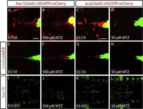
foxc1 acts upstream of acta2 in vSMC differentiation. A-D) Ablation of foxc1b and acta2 positive cells using Metronidazole (MTZ) from 3 to 4 dpf. Embryos are heterozygous for foxc1b- or acta2:Gal4 and UAS:NTR-mCherry. Ventral views show smooth muscle coverage along the ventral aorta in controls (A and C), but largely absent in MTZ treated embryos (B, D). E-F) Ventral views of mural cell marker mRNA (acta2 and pdgfrβ) in foxc1b:Gal4; UAS:NTR-mCherry embryos show a reduction in acta2 and increase in pdgfrβ in MTZ treated embryos (F) versus controls (E). G-H) Expression of acta2 is reduced in MTZ treated acta2:Gal4; UAS:NTR-mCherryembryos versus controls, while pdgfrβ expression is unchanged. I-L) Expression of foxc1b mRNA is reduced in MTZ treated foxc1b:Gal4; UAS:NTR-mCherry embryos compared to controls, while foxc1bmRNA is unchanged in controls and MTZ treated acta2:Gal4; UAS:NTR-mCherry embryos. Green hearts in A-H are due to the transgenesis marker (myl7:EGFP). Scale bar represents 50 μm. |
|
foxc1b expression is faithfully reported by the foxc1b:EGFP transgenic line in the zebrafish ventral head. A-F) 2 dpf and G-N) 4 dpf images of foxc1b expression in lateral (A-D, G-L) and ventral views (E, F, L-N). A) foxc1b mRNA (purple) is expressed in the aortic arches, jaw, ceratohyal, and periocular mesenchyme of the ventral head of the zebrafish embryo. B) foxc1b mRNA and foxc1b:EGFP transgene expression (brown) overlap (arrows) in the ceratohyal. C) foxc1b:EGFP transgene (green) is strongly expressed in the aortic arches and mesenchyme of the ventral head. D) foxc1b mRNA expression (green) overlaps with foxc1b:EGFP antibody stain (red) in the aortic arch arteries (arrows). E) Strong expression of foxc1b:EGFP in the ventral head including the ceratohyal. F) foxc1b mRNA expression overlaps with foxc1b:EGFP antibody stain in the ceratohyal. G) At 4 dpf, foxc1b mRNA is present in the aortic arch arteries, the epibranchial region of the ventral brain, ceratohyal, swim bladder, and intestine. H) foxc1b mRNA and foxc1b:EGFP overlap in cells surrounding the aortic arch arteries. I) foxc1b:EGFP is expressed in aortic arch arteries. J) foxc1b mRNA expression overlaps with foxc1b:EGFP antibody stain in the aortic arch arteries (arrows). K) egfp mRNA and foxc1b:EGFP overlap in cells surrounding the aortic arch arteries (arrows). L) foxc1b:EGFP is expressed around the ventral aorta and ceratohyal. M) foxc1b mRNA expression overlaps with foxc1b:EGFP antibody stain in the ventral aorta (arrows) and ceratohyal. N) egfp mRNA and foxc1b:EGFP overlap in cells surrounding the ventral aorta (arrows) and ceratohyal. Scale bars in A and G represent 200 µm. Scale bars in B and H represent 20 µm. Scale bars in C-F and I-N represent 50 µm. Insets in D, F, J, K, M and N show merged, foxc1b or egfp F-ISH (green), and foxc1b:EGFP (shown in red) antibody stain. AAA = aortic arch arteries, CH = ceratohyal, VA = ventral aorta. |
|
foxc1b:EGFP mRNA overlaps with acta2 expression in vascular smooth muscle cells. A-C) 2 dpf images and D-L) 4 dpf images. A) foxc1b mRNA expressing-cells are adjacent to kdrl:EGFP positive endothelium (brown, arrowheads). B) foxc1b mRNA (green) is detected adjacent to kdrl:EGFP antibody stain (red) in the aortic arch arteries. C) foxc1b mRNA is expressed in the ceratohyal. D) foxc1b mRNA expressing-cells are adjacent to the endothelium (arrowheads). E) foxc1b mRNA is in mural cells adjacent to kdrl:EGFP antibody stain (red) in the aortic arch arteries (arrowheads). F) foxc1b mRNA is observed in mural cells adjacent to the ventral aorta (arrowheads). G-L) At 4 dpf, foxc1b mRNA and acta2:EGFP (brown) are co-expressed in mural cells in the aortic arch arteries and brain vessels. Enlargements shown in J, K, and L. H) foxc1b mRNA is co-expressed with acta2:EGFP antibody stain (red) in the aortic arch arteries (arrowheads). I) foxc1b mRNA is co-expressed with acta2:EGFP around the ventral aorta (arrowheads). J) Enlargement of the ventral head in G shows overlap of foxc1b mRNA and acta2:EGFP on the aortic arch arteries (arrow). K) In the brain, there is overlap of foxc1b mRNA and acta2:EGFP (arrows). L) Enlargement of panel K, showing the overlap between foxc1b mRNA and acta2:EGFP, but also that foxc1b mRNA is expressed in smaller vessels distal to the termination point of acta2:EGFP (asterisks). Scale bars in A, D, J represent 20 µm. Scale bars in B, C, E, F, H, I represent 50 µm. Scale bar in G represents 100 µm. AAA = aortic arch arteries, CH = ceratohyal, VA = ventral aorta. |
|
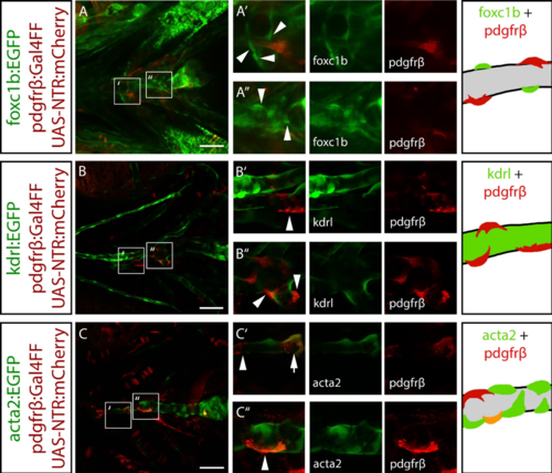
foxc1b:EGFP positive cells do not co-express a pericyte marker. A) foxc1b:EGFP is not co-expressed with pdgfrβ:mCherry in the ventral head. B) pdgfrβ:mCherry is associated with the endothelium (kdrl:EGFP) in the ventral head. C) pdgfrβ:mCherry and acta2:EGFP are partially co-expressed in ventral head mural cells. Schematics depict mural cell and endothelial marker expression that match the transgenes. Grey indicates presumptive endothelial patterns. Scale bars represent 50 µm. EXPRESSION / LABELING:
|
|
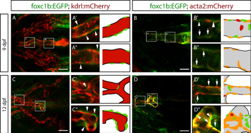
foxc1b:EGFP is expressed in vascular smooth muscle cells on the ventral aorta at larval stages. Ventral images of zebrafish embryos at 9 and 12 dpf. A) At 9 dpf, foxc1b:EGFP positive cells (arrowheads) are located adjacent to the kdrl:mCherry expressing endothelial cells of the ventral aorta (n = 4 embryos). B) At 9 dpf, foxc1b:EGFP and acta2:mCherry are co-expressed in mural cells (arrows) with the exception of a few single-positive cells (arrowheads; n = 4 embryos). C) At 12 dpf, foxc1b:EGFP positive cells continue to associate with endothelial cells of the ventral aorta (n = 6 embryos). D) At 12 dpf, most mural cells co-express foxc1b:EGFP and acta2:mCherry, although there are some single positive cells (n = 6 embryos). Grey colour in model indicates endothelial patterns. Scale bars represent 50 µm. |
|
foxc1b:EGFP positive cells in the trunk are associated with the endothelium and co-express acta2:mCherry at 4 dpf. Lateral imaging of dorsal aorta and intersegmental vessels in the trunk. A) foxc1b:EGFP positive cells are associated with the endothelium of both the dorsal aorta and the intersegmental vessels (arrowheads). B) foxc1b:EGFP positive cells co-express the smooth muscle marker acta2:mCherry on the dorsal aorta (arrows), but not on the intersegmental vessels (arrowhead). Scale bars represent 50 µm. |
|
Expression of new and known smooth muscle markers. Gene expression using colorimetric ISH in lateral view wholemount and sections, compared to endothelial (antibody staining for kdrl:EGFP, brown, column i), or smooth muscle (acta2:EGFP, brown, column ii) transgenes. Arrowheads depict gene expression associated with the endothelium, arrows depict gene expression co-expressed with smooth muscle. A) tagln, a known smooth muscle marker, is found in the aortic arch arteries, notochord tip, swim bladder, and gut in wholemount. In sections tagln is expressed in cells adjacent to the endothelium and is co-expressed with acta2:EGFP on the ventral aorta and aortic arch arteries. B) capn3b is expressed in cells adjacent to the endothelium in the brain. C) kcne4 is expressed adjacent to the endothelium and is co-expressed in smooth muscle on the ventral aorta and aortic arch arteries. D) pcolce2b is expressed adjacent to the endothelium and is co-expressed with acta2:EGFP. E) cnn1b is expressed adjacent to the endothelium of the aortic arch arteries. F) loxa is expressed solely in the smooth muscle of the bulbus arteriosus. G) lmod1b is expressed adjacent to the endothelium and overlaps with acta2:EGFP. H) desmb is expressed in a punctate pattern in the brain. I) myl9a is expressed adjacent to the endothelium, and partially overlaps with acta2:EGFP. J) tpm4b is expressed near the endothelium in the ventral head and is co-expressed with acta2:EGFP in the brain. K) si:ch211-247i17.1 is expressed near the endothelium and is co-expressed with acta2:EGFP. Scale bar for A-K (wholemount) represent 200 µm. Scale bars for sections (i and ii) represent 20 µm. |
|
foxc1a and foxc1b compound mutant morphology. Lateral images at 48 hpf (A) and 96 hpf (B), showing increasing cardiovascular insufficiency (pericardial edema, hydrocephalus) with progressive loss of foxc1a/foxc1b alleles. At 48 hpf, there is little morphological difference among all genotypes, except for mild edema in foxc1a mutants. At 96 hpf, there is severe edema in foxc1a-/- embryos, with the most severe phenotype with additional loss of foxc1b alleles. Scale bar represents 50 µm for all panels. |
|
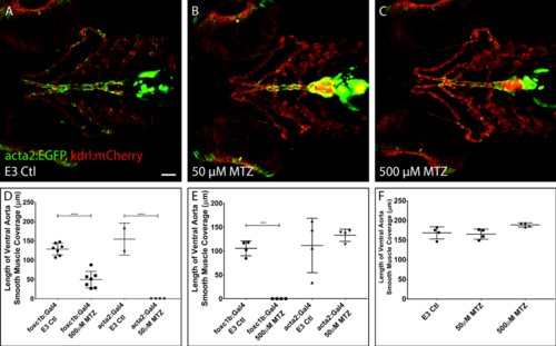
Treatment of Metronidazole (MTZ) does not affect vessel patterning or vSMC coverage along the ventral aorta in embryos. A-C) 4 dpf acta2:EGFP, kdrl:mCherry E3 control embryos lacking the NTR construct (A) show that treatment with MTZ does not affect vascular patterning or development of vSMC when using the same doses for acta2:Gal4 (B) or foxc1b:Gal4 (C) ablation. n = 4 embryos per panel. D) Quantification of vSMC coverage from data in Figure 9 A-D, using live transgenics (foxc1b:Gal4; UAS-NTR:mCherry or acta2:Gal4; UAS-NTR:mCherry) at 4 dpf. E) Quantification of vSMC coverage in Figure 9 E-H, using acta2 mRNA in situ hybridization. F) Quantification of vSMC coverage using live transgenics lacking NTR construct (acta2:EGFP, kdrl:mCherry). Data points in D-F represent individual embryos. *** = p < 0.001, ANOVA with Sidak’s multiple comparisons test. Scale bar represents 50 µm. |
Reprinted from Developmental Biology, 453(1), Whitesell, T.R., Chrystal, P.W., Ryu, J.R., Munsie, N., Grosse, A., French, C.R., Workentine, M.L., Li, R., Zhu, L.J., Waskiewicz, A., Lehmann, O.J., Lawson, N.D., Childs, S.J., foxc1 is required for embryonic head vascular smooth muscle differentiation in zebrafish, 34-47, Copyright (2019) with permission from Elsevier. Full text @ Dev. Biol.


