
IMAGE
Fig. 1
- ID
- ZDB-IMAGE-190823-22
- Genes
- Publication
- Whitesell et al., 2019 - foxc1 is required for embryonic head vascular smooth muscle differentiation in zebrafish
- All Figures
- Figures for Whitesell et al., 2019
Image
|
Figure Caption
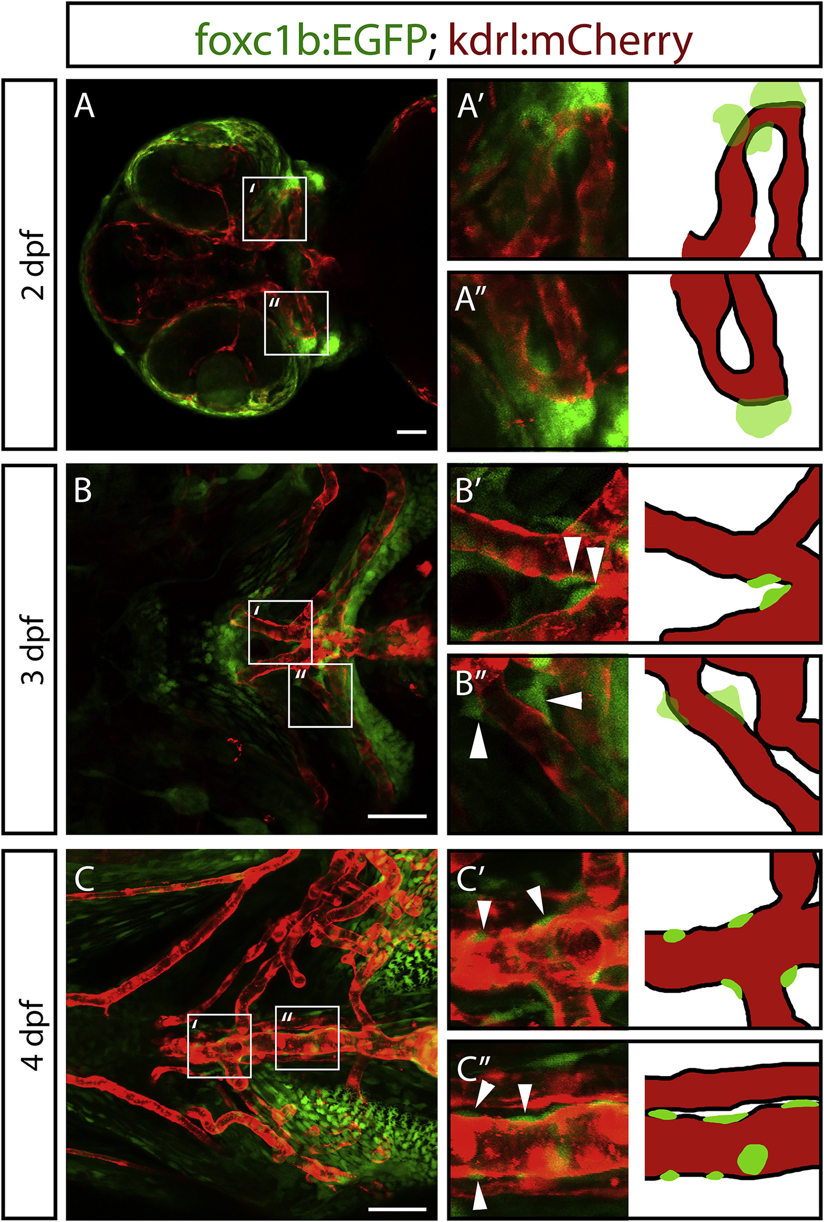
Fig. 1
foxc1b:EGFP expressing mesenchymal cells attach to vessels and take on a vSMC morphology. A) Images of the ventral head in 2 dpf zebrafish embryos show foxc1b:EGFP positive mesenchymal cells near the endothelium (kdrl:mCherry; n = 19 embryos). B) At 3 dpf, foxc1b:EGFP positive cells associate with the endothelium (n = 14 embryos). Arrowheads indicate mural cells associated with the endothelium. C) At 4 dpf, foxc1b:EGFP positive cells flatten and tightly associate with the endothelium (n = 11 embryos). Scale bars represent 50 μm.
Figure Data
Acknowledgments
This image is the copyrighted work of the attributed author or publisher, and
ZFIN has permission only to display this image to its users.
Additional permissions should be obtained from the applicable author or publisher of the image.
Reprinted from Developmental Biology, 453(1), Whitesell, T.R., Chrystal, P.W., Ryu, J.R., Munsie, N., Grosse, A., French, C.R., Workentine, M.L., Li, R., Zhu, L.J., Waskiewicz, A., Lehmann, O.J., Lawson, N.D., Childs, S.J., foxc1 is required for embryonic head vascular smooth muscle differentiation in zebrafish, 34-47, Copyright (2019) with permission from Elsevier. Full text @ Dev. Biol.