
- Title
-
Dlx3b/4b is required for early-born but not later-forming sensory hair cells during zebrafish inner ear development
- Authors
- Schwarzer, S., Spieß, S., Brand, M., Hans, S.
- Source
- Full text @ Biol. Open
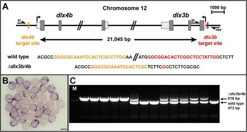
Generation of a dlx3b and dlx4b (dlx3b/4b) null allele. (A) Scheme of the dlx3b/4b bigene cluster at chromosome 12. Transcriptional start sites are indicated by arrows. Exon sequences with translated and untranslated regions are represented in dark and light grey, respectively. Positions of the CRISPR/Cas9 target sequences, separated by 21,045 bp, and their sequences in the wild-type and dlx3b/4b deletion allele are indicated in orange and red. Primers used for genotyping (1f, 2f and rev) are shown as half arrows. (B) In situ hybridization of dlx3b at early OEPD stages (three somites). Expression of dlx3b is absent in 25% of embryos obtained from a dlx3b/4b heterozygote incross. Scale bar: 500 µm. (C) Multiplex PCR using primers 1f, 2f and rev reveals the presence of the dlx3b/4b deletion (618 bp) and wild-type allele (473 bp). M indicates a marker for molecular size standard. |

Loss of Dlx3b/4b results in otic vesicle hypoplasia. (A-H) Live images of wild-type control embryos (A,E), dlx3b/4b mutants (B,F), foxi1 mutants (C,G) and dlx3b/4b; foxi1 double mutants (D,H) at 24 hpf (A-D) and 72 hpf (E-H). (I-L) Expression of starmaker (stm), a marker of the otic epithelium in wild-type control siblings (I), dlx3b/4b mutants (J), foxi1 mutants (K) and dlx3b/4b; foxi1 double mutants (L) at 24 hpf. Lateral views are seen with anterior to the left. Scale bars: 50 µm in H; 40 µm in L. EXPRESSION / LABELING:
PHENOTYPE:
|
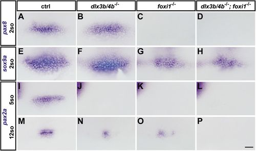
Loss of Dlx3b/4b results in compromised OEPD induction. In situ hybridization of pax8 (A-D), sox9a (E-H) and pax2a (I-P) at early OEPD (two and five somites) and placodal (12 somites) stages in wild-type control embryos (A,E,I,M), dlx3b/4b mutants (B,F,J,N), foxi1 mutants (C,G,K,O) and dlx3b/4b; foxi1 double mutants (D,H,L,P). (A-L) Dorsal views with anterior to the left. (M-P) Dorsolateral views with anterior to the left. Scale bar: 40 µm. EXPRESSION / LABELING:
PHENOTYPE:
|
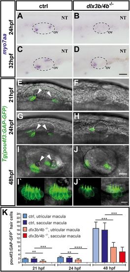
Delayed formation of sensory hair cells in dlx3b/4b-deficient embryos. (A-D) In comparison with control embryos at 24 hpf and 32 hpf, myo7aa expression is initially absent and subsequently restricted to the domain of the future utricular macula in dlx3b/4b mutants. (E-J) Expression of Tg(pou4f3:GAP-GFP) in control siblings and dlx3b/4b mutants at 21 hpf, 24 hpf and 48 hpf confirms delayed formation of sensory hair cells in dlx3b/4b-deficient embryos. Arrowheads indicate positions of the nascent otoliths in wild-type embryos. Note the initial onset of GFP in the prospective domain of the utricular macula and the subsequent expression in the future domain of the saccular macula in dlx3b/4b mutants. (I′,J′) Kinocilia formation appears normally in control siblings and dlx3b/4b mutants. Lateral views are shown with anterior to the left. NT, neural tube; OV, otic vesicle. Scale bars: 40 µm in D; 25 µm in J; 10 µm in J′. (K) Time course showing the mean number of pou4f3:GAP-GFP-positive hair cells in the domain of the future utricular and saccular domain of control (ctrl) and dlx3b/4b mutant embryos. dlx3b/4b-deficient embryos exhibit a significantly reduced number of sensory hair cells at 21 hpf and 24 hpf, and display significantly fewer hair cells at 48 hpf (**P<0,01; ***P<0,001; ****P<0,0001). Data are mean±s.e.m (n≥6 for each time point). EXPRESSION / LABELING:
PHENOTYPE:
|
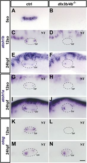
Analysis of upstream regulators of sensory hair cell and otolith development. (A-D) At OEPD (five somites) and placodal stages (12 somites), otic expression of atoh1b is absent in dlx3b/4b mutant embryos in comparison with wild-type siblings. (E,F) In contrast, atoh1b can be detected in dlx3b/4b mutants at 24 hpf although limited to the anterior portion of the otic vesicle. (G-J) Similarly, atoh1a is initially not expressed in the developing otic placode of dlx3b/4b-deficient embryos but present in the future domain of the utricular macula at 24 hpf. (K-N) Compared with wild-type control, otog expression is undetectable in dlx3b/4b mutants at placodal stages and restricted to the prospective domain of the utricular macula at 24 hpf. (A-D,G,H) Dorsolateral views with anterior to the left. (E,F,I,J) Lateral views with anterior to the left. OP, otic placode. Scale bar: 40 µm. EXPRESSION / LABELING:
PHENOTYPE:
|

OEPD-dependent neurogenesis in dlx3b/4b-deficient embryos. (A,B) Expression of neurod1 (blue) in control siblings and dlx3b/4b mutants at 24 hpf. Expression of stm (red) reveals the size reduction of the otic vesicle in dlx3b/4b mutants in comparison with wild-type embryos. (C,D) Expression of neurog1 in control and dlx3b/4b mutant embryos at 24 hpf. (E,F) Double staining of neurod1 (red) and tlx3b (blue) indicates increased production of anterior lateral line ganglion progenitors in dlx3b/4b mutants compared to control siblings at 24 hpf. (G,H) At 32 hpf, phox2a expression in dlx3b/4b mutants is indistinguishable from wild-type controls. (A,B,E,F) Lateral views with anterior to the left. (C,D,G,H) Dorsal views with anterior to the left. gALL, anterior lateral line ganglion progenitor; gVII, geniculate ganglion progenitor; gVIII, statoacoustic ganglion progenitor; gIX, petrosal ganglion progenitor; gX, nodose ganglion progenitor. Scale bar: 40 µm. |