Fig. 6
Fig. 6
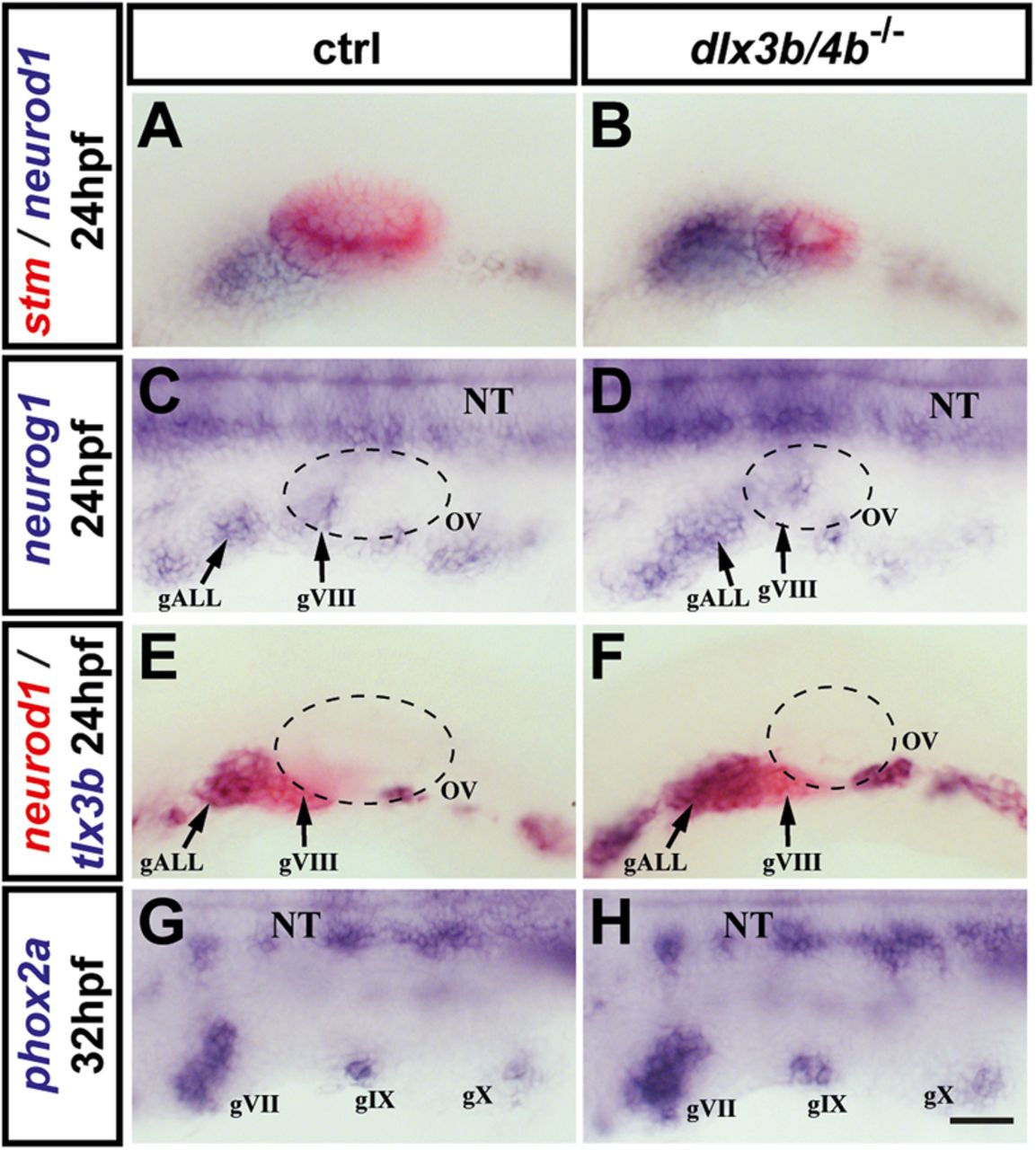
OEPD-dependent neurogenesis in dlx3b/4b-deficient embryos. (A,B) Expression of neurod1 (blue) in control siblings and dlx3b/4b mutants at 24 hpf. Expression of stm (red) reveals the size reduction of the otic vesicle in dlx3b/4b mutants in comparison with wild-type embryos. (C,D) Expression of neurog1 in control and dlx3b/4b mutant embryos at 24 hpf. (E,F) Double staining of neurod1 (red) and tlx3b (blue) indicates increased production of anterior lateral line ganglion progenitors in dlx3b/4b mutants compared to control siblings at 24 hpf. (G,H) At 32 hpf, phox2a expression in dlx3b/4b mutants is indistinguishable from wild-type controls. (A,B,E,F) Lateral views with anterior to the left. (C,D,G,H) Dorsal views with anterior to the left. gALL, anterior lateral line ganglion progenitor; gVII, geniculate ganglion progenitor; gVIII, statoacoustic ganglion progenitor; gIX, petrosal ganglion progenitor; gX, nodose ganglion progenitor. Scale bar: 40 µm.

