
- Title
-
Motor neuron-derived Thsd7a is essential for zebrafish vascular development via the Notch-dll4 signaling pathway
- Authors
- Liu, L.Y., Lin, M.H., Lai, Z.Y., Jiang, J.P., Huang, Y.C., Jao, L.E., Chuang, Y.J.
- Source
- Full text @ J. Biomed. Sci.

thsd7a was first expressed in primary motor neurons. Whole-mount ISH showed the spatiotemporal expression pattern of thsd7a, islet I and islet II at 24 hpf in wild type zebrafish embryos. Anterior is to the left. a: thsd7a transcripts were detected along the ventral edge of the neuron tube indicated by arrowheads. b, c: islet I (MiP and RoP marker) and islet II (CaP marker) were expressed in primary motor neurons along the ventral edge of the neuron tube indicated by arrowheads. Rohon-Beard sensory neurons can be seen along the dorsal edge of the neuron tube. a′, b′, c′: Enlarged images of the boxed regions in panel a, b and c, respectively. Scale bar is 100 µm EXPRESSION / LABELING:
|
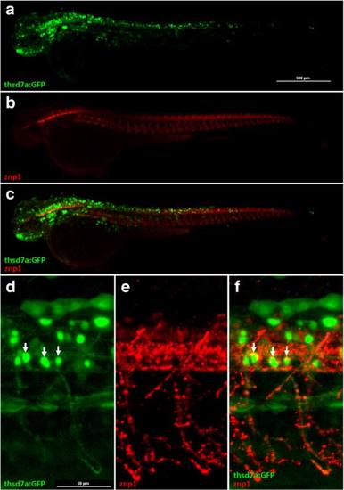
The signals of Tg(thsd7a:GFP) were detected in primary motor neurons. Representative images of the Tg(thsd7a:GFP) transgenic zebrafish at 48 hpf. Thsd7a and motor neurons were shown in green and red, respectively. Anterior is to the left. a-c GFP signals driven by thsd7a promoter were detected in the brain and the neural tube, consistent with ISH results. Moreover, the signals at the neural tube co-localized with anti-Znp1 antibody (shown in red). d-f Enlarged images of the neural tube showed GFP signals were expressed in primary motor neurons, indicated by arrows. EXPRESSION / LABELING:
|
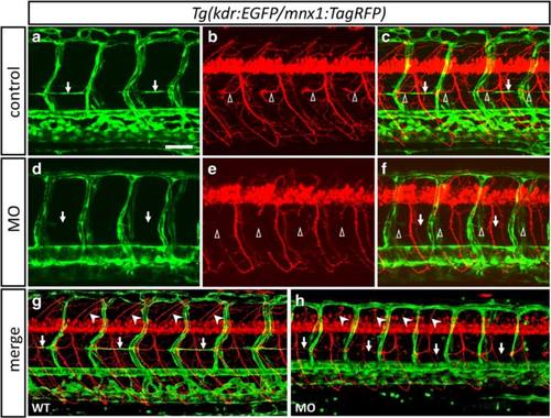
Loss of thsd7a impaired PAC, RoP axons and associated secondary motor neuron axons formation. Representative images showed the effect of Thsd7a knockdown on PAC angiogenesis and RoP neurogenesis by morpholino in Tg(kdr:EGFP/mnx1:TagRFP). Embryos were examined at 48 hpf (a-f) and 78 hpf (g-h). Anterior is to the left. a-c, g: Uninjected control. d-f, H: Embryos were injected with MO1 and MO2. Vasculature and motor neuron shown in green and red, respectively. PAC, RoP and MiP are indicated by arrows, open arrowheads and arrowheads, respectively. a, b: PAC and ROP axons were formed normally along HMS in control group. d: PAC was completely absent at HMS in morphants. e: RoP axons were absent at HMS. Moreover, MiP axons and some secondary motoneuron grew aberrantly. c, f: Merged images of panel a, b and d, e respectively. g: PAC and primary motoneurons grew normally. h: Some initially absent RoP axons reappeared but their trajectory were slightly shifted and PAC still absent. MiP axons were shorter with abberant outgrowth. Scale bar is 100 µm. EXPRESSION / LABELING:
PHENOTYPE:
|

ZFIN is incorporating published figure images and captions as part of an ongoing project. Figures from some publications have not yet been curated, or are not available for display because of copyright restrictions. |

Thsd7a knockdown resulted in aberrant Notch1b expression pattern. Whole-mount ISH revealed the spatiotemporal expression patterns of notch1b at 48 hpf with either Thsd7a MO1 (a′-c′) or 5msMO1 (a-c). In the control group, notch1b transcripts were detected in the hind brain with a grid-like pattern (a). In comparison, Thsd7a morphants lost the grid-like expression pattern of notch1b (a′), which were indicated by the white arrows. Reduced expression of notch1b in the spinal cord (b′) was observed. In Thsd7a morphants, notch1b ectopic expression was observed throughout the endocardium in the heart tube (c′) indicated by white arrow head |
|
ZFIN is incorporating published figure images and captions as part of an ongoing project. Figures from some publications have not yet been curated, or are not available for display because of copyright restrictions. |
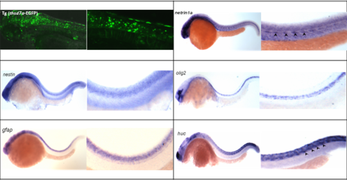
Comprehensive in situ hybridization analysis of thsd7a expression to different neuronal markers. The expression of different neuronal markers include nestin (progenitor cells of nervous system), olig2 (oligodendrocyte), gfap (glial fibrillary acidic protein; astrocyte), huc (Hu antigen C; mature neuron), islet I (MiP and RoP motoneuron), islet II (CaP motoneuron) and netrin1a (neuron tube and HMS). |
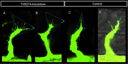
The tip cells on ISV showed protrusions without specific orientation in Thsd7a knockdown zebrafsih. Representative images showed the effects of Thsd7a knockdown by injecting Thsd7a MO2 into Tg(fli1:EGFP) zebrafish embryos; 5msMO2 was used as control. The morphants were then observed at 27 ~ 34hpf. In the control group, the tip cell on ISV displayed tree-shape morphology with single main protrusion (A and B). After knockdown of Thsd7a, the tip cell displayed fan-shape morphology with disorientation (C and D). Scale bar is 10 µm. |