
Fig. 1
- ID
- ZDB-IMAGE-160908-11
- Genes
- Publication
- Liu et al., 2016 - Motor neuron-derived Thsd7a is essential for zebrafish vascular development via the Notch-dll4 signaling pathway
- All Figures
- Figures for Liu et al., 2016
|
Fig. 1
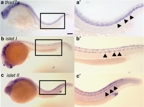
thsd7a was first expressed in primary motor neurons. Whole-mount ISH showed the spatiotemporal expression pattern of thsd7a, islet I and islet II at 24 hpf in wild type zebrafish embryos. Anterior is to the left. a: thsd7a transcripts were detected along the ventral edge of the neuron tube indicated by arrowheads. b, c: islet I (MiP and RoP marker) and islet II (CaP marker) were expressed in primary motor neurons along the ventral edge of the neuron tube indicated by arrowheads. Rohon-Beard sensory neurons can be seen along the dorsal edge of the neuron tube. a′, b′, c′: Enlarged images of the boxed regions in panel a, b and c, respectively. Scale bar is 100 µm