Image
Figure Caption
Fig. 2
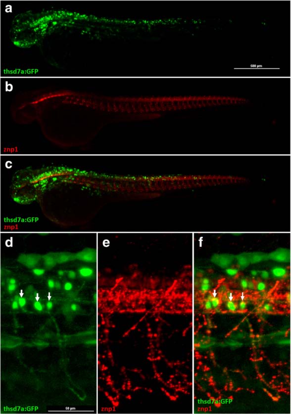
The signals of Tg(thsd7a:GFP) were detected in primary motor neurons. Representative images of the Tg(thsd7a:GFP) transgenic zebrafish at 48 hpf. Thsd7a and motor neurons were shown in green and red, respectively. Anterior is to the left. a-c GFP signals driven by thsd7a promoter were detected in the brain and the neural tube, consistent with ISH results. Moreover, the signals at the neural tube co-localized with anti-Znp1 antibody (shown in red). d-f Enlarged images of the neural tube showed GFP signals were expressed in primary motor neurons, indicated by arrows.
Figure Data
Acknowledgments
This image is the copyrighted work of the attributed author or publisher, and
ZFIN has permission only to display this image to its users.
Additional permissions should be obtained from the applicable author or publisher of the image.
Full text @ J. Biomed. Sci.

