- Title
-
Lxr regulates lipid metabolic and visual perception pathways during zebrafish development
- Authors
- Pinto, C.L., Kalasekar, S.M., McCollum, C.W., Riu, A., Jonsson, P., Lopez, J., Swindell, E., Bouhlatouf, A., Balaguer, P., Bondesson, M., Gustafsson, J.Å.
- Source
- Full text @ Mol. Cell. Endocrinol.
|
The LXR ligands T0901317 and GW3965 activate zfLxr and affect gene expression in zebrafish larvae. Transactivation assays performed in HG5LN cells stably transfected with chimeric GAL4-zfLxr LBD in the presence of increasing concentrations of T0901317 and GW3965. Results are presented as mean ± s.d. of 3 independent experiments. (A) Heat map of differentially expressed genes in 6 dpf zebrafish larvae exposed to T0901317 or GW3965 as determined by microarray analysis (absolute fold change e |±1.4|, FDR-adjusted P≤0.05) (B). Venn diagram illustrating the number of statistically significant genes that were co-regulated in zebrafish larvae treated with T0901317 or GW3965 (C). |

ZFIN is incorporating published figure images and captions as part of an ongoing project. Figures from some publications have not yet been curated, or are not available for display because of copyright restrictions. |

ZFIN is incorporating published figure images and captions as part of an ongoing project. Figures from some publications have not yet been curated, or are not available for display because of copyright restrictions. |
|
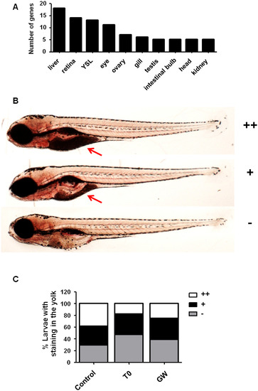
Increased yolk consumption in zebrafish larvae exposed to the Lxr ligands. Tissue enrichment analysis predicted the liver, retina/eye and yolk syncytial layer (YSL) to be affected by exposure of zebrafish larvae to the Lxr ligands (A). Zebrafish larvae were scored for Oil Red O (ORO) staining in the yolk region from no staining (), mild (+) to strong (++) staining (B). Number of zebrafish larvae exposed from 4 dpf to 6 dpf to 0.1% DMSO (vehicle), 2 µM T0 and 1 µM GW presenting different staining intensities in the yolk (n = 150 larvae for control, n = 148 for T0 and n = 150 for GW) (C). Red arrow indicates the yolk. (For interpretation of the references to colour in this figure legend, the reader is referred to the web version of this article.) PHENOTYPE:
|
|
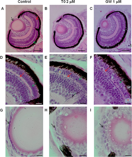
Morphological alterations in the eye of zebrafish larvae exposed to the Lxr ligands. Hematoxylin and eosin staining of eye sections of zebrafish larvae exposed to vehicle only (0.1% DMSO), 2 µM T0 and 1 µM GW from 6 hpf to 4 dpf, showing abnormalities in the lens and retina (A–I). Black arrow indicates lens epithelium red arrow indicates outer plexiform layer and green arrow indicates photoreceptor layer. Disorganized outer plexiform and photoreceptor layers with indications of photoreceptor cell death (indicated by red asterisks) were observed in zebrafish exposed to T0 (B, E) and GW (C, F) compared to control (A, D). The lens epithelial cell layer shows abnormal arrangement and morphology in zebrafish larvae treated with T0 (B, H) and GW (C, I). (Scale bars: A–C: 50 µm: D–I: 10 µm). (For interpretation of the references to colour in this figure legend, the reader is referred to the web version of this article.) PHENOTYPE:
|
|
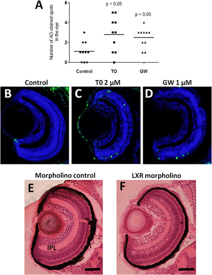
Apoptosis and morphological alterations in the eyes of Lxr ligand-treated or Lxr morpholino injected zebrafish larvae. Number of acridine orange stained spots in eyes of 4 dpf zebrafish larvae treated with T0 and GW (A). TUNEL staining of eye sections of zebrafish larvae treated vehicle only (B), T0 (C) or GW (D), Hematoxylin and eosin staining of eye sections of control morpholino (E) and Lxr morpholino (F) zebrafish larvae at 4 dpf. Representative images are shown. IPL; inner plexiform layer (Scale bars 50 µm). (For interpretation of the references to colour in this figure legend, the reader is referred to the web version of this article.) PHENOTYPE:
|

ZFIN is incorporating published figure images and captions as part of an ongoing project. Figures from some publications have not yet been curated, or are not available for display because of copyright restrictions. EXPRESSION / LABELING:
PHENOTYPE:
|

ZFIN is incorporating published figure images and captions as part of an ongoing project. Figures from some publications have not yet been curated, or are not available for display because of copyright restrictions. EXPRESSION / LABELING:
PHENOTYPE:
|

ZFIN is incorporating published figure images and captions as part of an ongoing project. Figures from some publications have not yet been curated, or are not available for display because of copyright restrictions. |
Reprinted from Molecular and Cellular Endocrinology, 419, Pinto, C.L., Kalasekar, S.M., McCollum, C.W., Riu, A., Jonsson, P., Lopez, J., Swindell, E., Bouhlatouf, A., Balaguer, P., Bondesson, M., Gustafsson, J.Å., Lxr regulates lipid metabolic and visual perception pathways during zebrafish development, 29-43, Copyright (2016) with permission from Elsevier. Full text @ Mol. Cell. Endocrinol.


