
Fig. 5
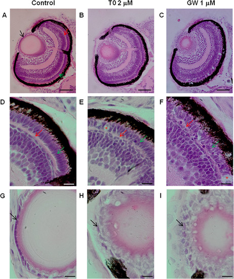
Morphological alterations in the eye of zebrafish larvae exposed to the Lxr ligands. Hematoxylin and eosin staining of eye sections of zebrafish larvae exposed to vehicle only (0.1% DMSO), 2 µM T0 and 1 µM GW from 6 hpf to 4 dpf, showing abnormalities in the lens and retina (A–I). Black arrow indicates lens epithelium red arrow indicates outer plexiform layer and green arrow indicates photoreceptor layer. Disorganized outer plexiform and photoreceptor layers with indications of photoreceptor cell death (indicated by red asterisks) were observed in zebrafish exposed to T0 (B, E) and GW (C, F) compared to control (A, D). The lens epithelial cell layer shows abnormal arrangement and morphology in zebrafish larvae treated with T0 (B, H) and GW (C, I). (Scale bars: A–C: 50 µm: D–I: 10 µm). (For interpretation of the references to colour in this figure legend, the reader is referred to the web version of this article.) |
| Fish: | |
|---|---|
| Condition: | |
| Observed In: | |
| Stage: | Day 4 |
Reprinted from Molecular and Cellular Endocrinology, 419, Pinto, C.L., Kalasekar, S.M., McCollum, C.W., Riu, A., Jonsson, P., Lopez, J., Swindell, E., Bouhlatouf, A., Balaguer, P., Bondesson, M., Gustafsson, J.Å., Lxr regulates lipid metabolic and visual perception pathways during zebrafish development, 29-43, Copyright (2016) with permission from Elsevier. Full text @ Mol. Cell. Endocrinol.