
Fig. 4
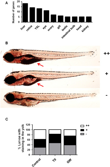
Increased yolk consumption in zebrafish larvae exposed to the Lxr ligands. Tissue enrichment analysis predicted the liver, retina/eye and yolk syncytial layer (YSL) to be affected by exposure of zebrafish larvae to the Lxr ligands (A). Zebrafish larvae were scored for Oil Red O (ORO) staining in the yolk region from no staining (), mild (+) to strong (++) staining (B). Number of zebrafish larvae exposed from 4 dpf to 6 dpf to 0.1% DMSO (vehicle), 2 µM T0 and 1 µM GW presenting different staining intensities in the yolk (n = 150 larvae for control, n = 148 for T0 and n = 150 for GW) (C). Red arrow indicates the yolk. (For interpretation of the references to colour in this figure legend, the reader is referred to the web version of this article.) |
| Fish: | |
|---|---|
| Condition: | |
| Observed In: | |
| Stage: | Day 6 |
Reprinted from Molecular and Cellular Endocrinology, 419, Pinto, C.L., Kalasekar, S.M., McCollum, C.W., Riu, A., Jonsson, P., Lopez, J., Swindell, E., Bouhlatouf, A., Balaguer, P., Bondesson, M., Gustafsson, J.Å., Lxr regulates lipid metabolic and visual perception pathways during zebrafish development, 29-43, Copyright (2016) with permission from Elsevier. Full text @ Mol. Cell. Endocrinol.