- Title
-
C1q-like inhibits p53-mediated apoptosis and controls normal hematopoiesis during zebrafish embryogenesis
- Authors
- Mei, J., Zhang, Q.Y., Li, Z., Lin, S., and Gui, J.F.
- Source
- Full text @ Dev. Biol.
|
Molecular characterization and expression pattern of zebrafish C1q-like. (A) Amino-acid alignment of C1q-like from zebrafish with putative C1q-like factor from Carassius auratus (Ca), brook trout (Salvelinus fontinalis, Sf), rainbow trout (Oncorhynchus mykiss, Om), Norway rat (Rattus norvegicus, Rn) and rhesus monkey (Macaca mulatta, Mm). Similar and identical amino acids are highlighted in grey and black boxes. And the percentages of identities and similarities were shown compared zebrafish C1q-like with other C1q-like factors. (B, C) Expression of C1q-like in adult tissues (B) and embryonic development stages (C) evaluated by real-time PCR and normalized to beta-actin mRNA. The data represent mean ± SD of three separate experiments. (D) Expression pattern of zebrafish C1q-like. Whole-mount RNA in situ hybridization and immunohistochemistry were performed using a C1q-like specific antisense riboprobe (a, c, e, g) and anti-C1q-like antibody (b, d, f, h) on embryos at the indicated stages. The arrows indicated the signals in the anterior and head. Embryos in panels a and b are lateral views with the animal pole toward the top. The embryos in other panels are lateral views, with dorsal toward the top and anterior to the left. |
|
Effect of the C1q-like knockdown on embryo development. (A) Embryo phenotypes observed under the microscope (a, c, e) and the head phenotypes stained with Alcian Blue (b, d, f). The arrows show the defected craniofacial phenotypes in the C1q-like morphant. Abbreviations: cb(I–V), first to fifth ceratobranchial; ch, ceratohyal; mk, Meckel's cartilage; pf, pectoral fin; pq, palatoquandrate. Each picture represents typical results out of three separate experiments (30 embryos in each experiment). (B, C) The statistical data of three independent experiments on C1q-like knockdown and C1q-like over-expression (B), as well as C1q-like mRNA rescue and p53 MO co-injection (B). Results are represented as mean ± SD of three separate experiments (60 embryos in each experiment). *p < 0.05. (D) Western blot detection of C1q-like knockdown during embryogenesis. The protein extracts from 30 embryos (28 hpf and 32 hpf) were analyzed by Western blot using the polyclonal anti-C1q-like antibody. A band of about 24 KD was not detected in C1q-like morphants. The picture represents typical result from three separate experiments. |
|
Developmental defects and caspase 3/9 activity changes in the C1q-like MO embryos. (A) The head phenotypes revealed by AO staining (a, c, e, g, i) and TUNEL assays (b, d, f, h, j). The pictures are representative of at least three experiments (more than 30 embryos in each experiment). (B) The tail phenotypes revealed by AO staining (a, c, e, g, i) and TUNEL assays (b, d, f, h, j). The picture a2–j2 showing the corresponding amplification signals of the apoptotic cells in intermediate cell mass (ICM) region from a1–j1. (C, D) Caspase-9 (C) and 3 (D) activity changes in the shown experiments. The activities were measured from embryos at 28 hpf by cleavage of AFC from synthetic substrate, were expressed as change in FAU/μg protein/h. The data represent mean ± SD of three separate experiments from thirty injected embryos. *p < 0.05. PHENOTYPE:
|
|
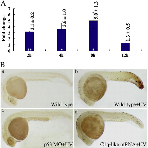
Developmental effects of C1q-like over-expression and UV exposure on zebrafish embryo development. (A) C1q-like expression changes detected by Q-RT-PCR at different times after the embryo exposure to UV. The ratio of C1q-like to β-actin in untreated embryos was set to 1 in every stage, and all UV treated embryos were normalized relative to this value. The data represent mean ± SD of three separate experiments. *p < 0.05 and **p < 0.01. (B) Normal phenotype of control wild-type embryo (a) and the UV exposed embryo phenotypes revealed by TUNEL assays in wild-type (b), p53 MO (c) and C1q-like mRNA injected (d) embryos. Each picture represents typical result from three experiments (more than 20 embryos in each experiment). PHENOTYPE:
|
|
Cell-cycle-progression defects in the C1q-like morphant embryos. (A) The up-regulated expression changes of p21WAF1/CIP-1 (p21), p53, mdm2, bax and caspase 3 revealed by Q-RT-PCR in the C1q-like morphants. The ratio of these genes to β-actin in control embryos was set to 1, and all C1q-like MO treated embryos were normalized relative to this value. The date represent mean ± SD of three separate experiments. (B) The labeled G2/M phase cells of head regions in control (a), the C1q-like morphant (b) and co-knockdowns of C1q-like and p53 (c) embryos at 24 hpf recognized with the antibody against phosphorylated histone H3 (pH3). (C) A typical FACS analysis data of propidium-iodide-labeled cells in control (a), the C1q-like morphant (b) and co-knockdowns of C1q-like and p53 (c) embryos at 24 hpf. (D) The statistical data of three independent experiments of (C). *p < 0.05 and **p < 0.01. |
|
The effects of C1q-like knockdown on primitive hematopoiesis. (A) Whole in situ hybridization of three hematopoietic marker genes gata-1 (a, b), scl (c, d) and hbae1 (e, f) in control MO embryos (a, c, e) and in the C1q-like morphant embryos (b, d, f) at 24 hpf. (B) Whole in situ hybridization of the hematopoietic marker gene hbae1 in control MO embryos (a, b) and in the C1q-like morphant embryos (c, d) at 48 hbf. The hybridization experiments were performed for 3 times, and at least 30 embryos were used for each experiment. The embryos are viewed at lateral views (a, c) and ventral views (b, d). The arrows indicate in situ hybridization signal. (C) Q-RT-PCR analysis of the expression changes of scl, gata-1 and hbae1 in 24 hpf and 48 hpf embryos injected with control MO or C1q-like MO. Results represent mean ± SD of three separate experiments. **p < 0.01 and ***p < 0.001. |
|
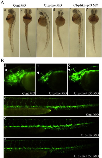
C1q-like MO affects the hematopoiesis and brain blood vessel organization. (A) The embryo phenotypes stained by O-dianisidine at 48 hpf. The embryos are shown as lateral views (a, c, e) and ventral views. (B) The effect of C1q-like MO on brain (a, b, c) and tail (d, e, f) blood vessels in the flk-1:GFP-transgenic zebrafish. The blood vessels of embryos were imaged at 48 hpf at 10x. Arrows show the mesencephalic vein (MsV). Each picture represents typical results from three experiments (more than 20 embryos in each experiment). |
|
Fig. 9. C1q-like is essential for defense and survival in response to bacterial infection. (A) C1q-like expression changes detected by Q-RT-PCR at different times after the embryos infected by the pathogen bacterium Aeromonas hydrophila at 24 hpf. The ratio of C1q-like to β-actin in untreated embryos was set to 1 in every stage, and all infected embryos were normalized relative to this value. The data represent mean ± SD of three separate experiments. Hpi means hours post infection. (B) At 24 hpf, the normal control wild-type embryos, the over-expressed C1q-like embryos, and the C1q-like morphant embryos were respectively challenged with the pathogen bacterium Aeromonas hydrophila, and the cumulative mortality were recorded at the indicated times after infection. The data represents mean ± SD of three separate experiments. |
|
The effect of C1q-like MO to midbrain–hindbrain specification. In situ hybridization of pax2.1, fgf8, her5, epha4a and krox-20 showed the distributions of these genes in Cont MO and C1q-like MO-injected embryos. Each picture represents typical results out of three experiments (20 embryos in each experiment). EXPRESSION / LABELING:
|

Unillustrated author statements PHENOTYPE:
|
Reprinted from Developmental Biology, 319(2), Mei, J., Zhang, Q.Y., Li, Z., Lin, S., and Gui, J.F., C1q-like inhibits p53-mediated apoptosis and controls normal hematopoiesis during zebrafish embryogenesis, 273-284, Copyright (2008) with permission from Elsevier. Full text @ Dev. Biol.


 p < 0.05.
