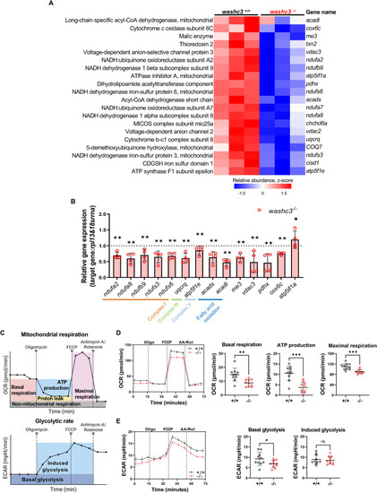
Loss of Washc3 results in suppression of mitochondrial proteins and mRNA leading to decreased mitochondrial respiration in adult zebrafish heart. (A) Heatmap of mitochondria related proteins significantly decreased in the washc3 mutant hearts compared to washc3+/+ at 1 year old. (B) qRT-PCR analysis and relative expression of listed genes from (A) in adult zebrafish heart (SD, n = 4, Mann–Whitney test, **p < 0.005 vs. washc3+/+). (C) Descriptive illustration of seahorse assay (XF Mito Stress assay) (created by Biorender.com). Oxygen consumption rate (OCR) and extracellular acidification rate (ECAR) are measured before and after treatment of Oligomycin, FCCP, and AA/Rot. Each colored boxes represent the states of respiration possibly read out from the OCR or the ECAR curves. (D,E) OCR and ECAR curves in response to washc3+/+ and washc3−/− primary cardiomyocytes dissociated from 1 year old adult zebrafish heart (SEM, n = 10). Corresponding quantifications of fundamental parameters shows significant decrease of basal respiration, ATP production, maximal respiration, basal glycolysis, and induced glycolysis in washc3−/− primary cardiomyocytes (SD, n = 10 well per group, Mann–Whitney test, ns = not significant, * p < 0.05, **p < 0.005, ***p < 0.001 vs. washc3+/+ cardiomyocytes).
|
