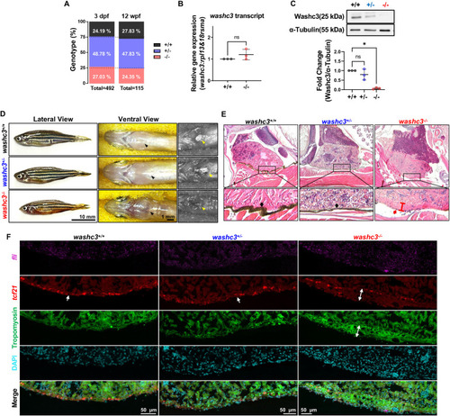
Aged Washc3 knockout zebrafish exhibits phenotypes of pericardial agenesis and compact outer layer of ventricle. (A) Relative ratio of offspring at 3 dpf and 12 weeks post fertilization (wpf) from washc3+/− adult zebrafish (3 dpf: washc3+/+, n = 119; washc3+/−, n = 240; washc3−/−, n = 133; total n = 492; 12 wpf: washc3+/+, n = 32; washc3+/−, n = 55; washc3−/−, n = 28; total n = 115). (B) qRT-PCR analysis of washc3 transcript at 1 year old adult zebrafish hearts (SD, n = 3, Mann–Whitney test, ns = not significant vs. washc3+/+). (C) Western blot analysis of Washc3 and α-Tubulin (SD, n = 3, Kruskal–Wallis test, *p < 0.05 vs. washc3+/+). Washc3 protein level was lost in the heart of 1 year old washc3−/−. (D) Representative lateral (left panel) and ventral (right panel) view of 17-months-old siblings (washc3+/+, washc3+/−) and washc3−/− zebrafish. Black arrows on the ventral view marks the position of the heart before incision. Yellow arrows on the right panel of ventral view denotes pericardial membrane and its location after incision (Scale bar: Lateral view: 10 mm; Ventral view, 1 mm). (E) Hematoxylin and Eosin staining (H&E) staining of paraffin sections prepared from the whole mount aged zebrafish. Bottom panels are enlarged images from squared regions of top panels. Black arrows indicate pericardial membranes, and red arrow denotes outer layer of ventricle in washc3−/−. In the heart of washc3−/−, pericardial layer is not observed and outer layer of ventricle is thicker compared to siblings (washc3+/+, washc3+/−). (G) Confocal images of RNA scope staining with fli (endocardial cell: magenta) and tcf21 (epicardial cell: red), as well as IF staining with Tropomyosin (myocardial cell: green) and DAPI (cyan) (scale bar: 50 μm). Outer layer of ventricle in washc3−/− is thickened where tcf21+ cells are widely dispersed in the compact myocardium.
|
