FIGURE

Figure 6

ID
ZDB-FIG-260311-692
Publication
Kim et al., 2026 - WASHC3 knockout disrupts mitochondrial protein homeostasis and energy metabolism in cardiomyocytes
Other Figures
All Figure Page
Back to All Figure Page
Figure 6

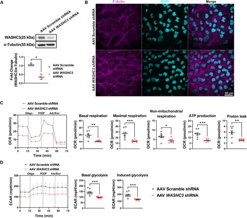
AAV-mediated knockdown of WASHC3 leads to defective mitochondrial respiration in human ventricular cardiomyocytes (AC16). (A) Immunoblot of WASHC3 and α-Tubulin (loading control) using AC16 cell lysate transduced with Adeno-associated virus (AAV) harboring scramble or WASHC3 shRNA (SD, n = 4; Mann–Whitney test, *p<0.05 vs. AAV6-Scramble-shRNA). (B) Confocal images of AC16 cells stained with F-Actin (magenta) and DAPI (cyan) at 72 h after transduction (scale bar: 50 μm). (C,D)) OCR and ECAR curves in AAV-scramble shRNA or -WASHC3 shRNA transduced AC16 cells (mean ± SEM, n = 10 well per group). Correspondent quantitative analyses of parameters reveal significantly repressed of mitochondrial respiration and glycolytic activity by AAV-mediated WASHC3 knockdown. Basal respiration, Maximal respiration, non-mitochondrial respiration, ATP production, proton leak, basal glycolysis, and induced glycolysis were showed. (SD, n = 10 well per group of AC16 cells, Mann–Whitney test, *p<0.05, **p<0.005, ***p<0.001 vs. Scramble shRNA).

Expression Data

Expression Detail
Antibody Labeling
Phenotype Data

Phenotype Detail
Acknowledgments
This image is the copyrighted work of the attributed author or publisher, and ZFIN has permission only to display this image to its users. Additional permissions should be obtained from the applicable author or publisher of the image. Full text @ Front Cardiovasc Med