
Fig. 3 - Supplemental 1
- ID
- ZDB-FIG-260217-12
- Publication
- Chiu et al., 2026 - Glial betaPix is essential for blood vessel development in the zebrafish brain
- Other Figures
- All Figure Page
- Back to All Figure Page
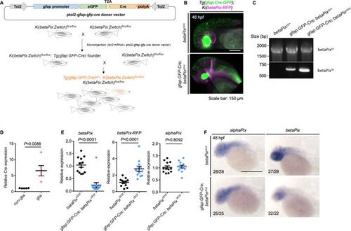
Generating glial-specific betaPix knockout zebrafish. (A) Schematic diagram illustrates establishment of glial-specific betaPix knockout zebrafish. (B) 3D reconstruction of the heads at 48 hpf with lateral view, anterior to left. Glial-specific Cre expression (green) was only shown in gfap:GFP-Cre; betaPixct/ct mutant embryos, which overlaps with betaPix:RFP expression (magenta). (C) Genomic PCR analysis of the betaPixct- or betaPixm-unique sequences in betaPixct/ct siblings, gfap:GFP-Cre; betaPixct/+ heterozygous mutants, and gfap:GFP-Cre; betaPixct/ct homozygous mutants. (D) qRT-PCR analysis revealing Cre expression in gfap:GFP-positive glial population and gfap:GFP-negative non-glial population that were sorted from gfap:GFP-Cre; betaPixct/ct embryos. Each dot represents one embryo. Data are presented in mean ± SEM; unpaired Student’s t-test with individual p-values mentioned in the figure. (E) qRT-PCR analysis showing betaPix, betaPix-RFP, and alphaPix expression in betaPixct/ct control siblings and gfap:GFP-Cre; betaPixct/ct mutant embryos at 48 hpf. Each dot represents one embryo. Data are presented in mean ± SEM; unpaired Student’s t-test with individual p-values mentioned in the figure. (F) Whole-mount RNA in situ hybridization revealing that alphaPix was comparable but betaPix decreased in gfap:GFP-Cre; betaPixct/ct mutants compared with betaPixct/ct siblings at 48 hpf. Lateral view with anterior to the left. Individual scale bars are indicated in the figure. |